

Sensorimotor Oscillations in Human Infants during an Innate Rhythmic Movement
- PMID: 38672051
- PMCID: PMC11047852
- DOI: 10.3390/brainsci14040402
Sensorimotor Oscillations in Human Infants during an Innate Rhythmic Movement
Abstract
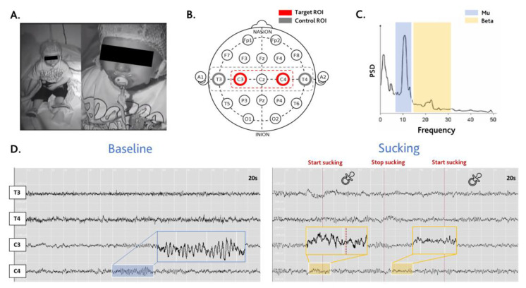
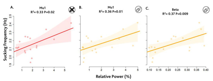
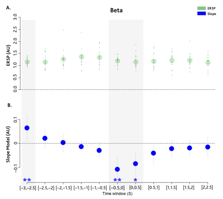
The relationship between cerebral rhythms and early sensorimotor development is not clear. In recent decades, evidence revealed a rhythmic modulation involving sensorimotor processing. A widely corroborated functional role of oscillatory activity is to coordinate the information flow across sensorimotor networks. Their activity is coordinated by event-related synchronisation and desynchronisation in different sensorimotor rhythms, which indicate parallel processes may be occurring in the neuronal network during movement. To date, the dynamics of these brain oscillations and early sensorimotor development are unexplored. Our study investigates the relationship between the cerebral rhythms using EEG and a typical rhythmic movement of infants, the non-nutritive sucking (NNS) behaviour. NNS is an endogenous behaviour that originates from the suck central pattern generator in the brainstem. We find, in 17 infants, that sucking frequency correlates with beta synchronisation within the sensorimotor area in two phases: one strongly anticipating (~3 s) and the other encompassing the start of the motion. These findings suggest that a beta synchronisation of the sensorimotor cortex may influence the sensorimotor dynamics of NNS activity. Our results reveal the importance of rapid brain oscillations in infants and the role of beta synchronisation and their possible role in the communication between cortical and deep generators.
Keywords: beta synchronisation; brain oscillations; infants; non-nutritive sucking; sensorimotor.
Conflict of interest statement
The authors declare no conflicts of interest.
Figures
Similar articles
-
Dynamic modulation of cortico-muscular coupling during real and imagined sensorimotor synchronisation.Neuroimage. 2021 Sep;238:118209. doi: 10.1016/j.neuroimage.2021.118209. Epub 2021 May 26. Neuroimage. 2021. PMID: 34051354
-
Abnormal Nutritive Sucking as an Indicator of Neonatal Brain Injury.Front Pediatr. 2021 Jan 12;8:599633. doi: 10.3389/fped.2020.599633. eCollection 2020. Front Pediatr. 2021. PMID: 33511093 Free PMC article. Review.
-
Lateralized alpha-band cortical networks regulate volitional modulation of beta-band sensorimotor oscillations.Neuroimage. 2014 Feb 15;87:147-53. doi: 10.1016/j.neuroimage.2013.10.003. Epub 2013 Oct 10. Neuroimage. 2014. PMID: 24121086
-
EEG Oscillations Are Modulated in Different Behavior-Related Networks during Rhythmic Finger Movements.J Neurosci. 2016 Nov 16;36(46):11671-11681. doi: 10.1523/JNEUROSCI.1739-16.2016. J Neurosci. 2016. PMID: 27852775 Free PMC article.
-
The ups and downs of β oscillations in sensorimotor cortex.Exp Neurol. 2013 Jul;245:15-26. doi: 10.1016/j.expneurol.2012.09.014. Epub 2012 Sep 27. Exp Neurol. 2013. PMID: 23022918 Review.
References
Grants and funding
LinkOut - more resources
Full Text Sources
