. 2024 Mar 23;12(4):717.
doi: 10.3390/biomedicines12040717.

NMDA Receptor Antagonist Memantine Ameliorates Experimental Autoimmune Encephalomyelitis in Aged Rats

Affiliations

NMDA Receptor Antagonist Memantine Ameliorates Experimental Autoimmune Encephalomyelitis in Aged Rats

Biljana Bufan et al. Biomedicines. .

Abstract

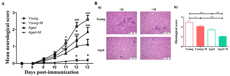
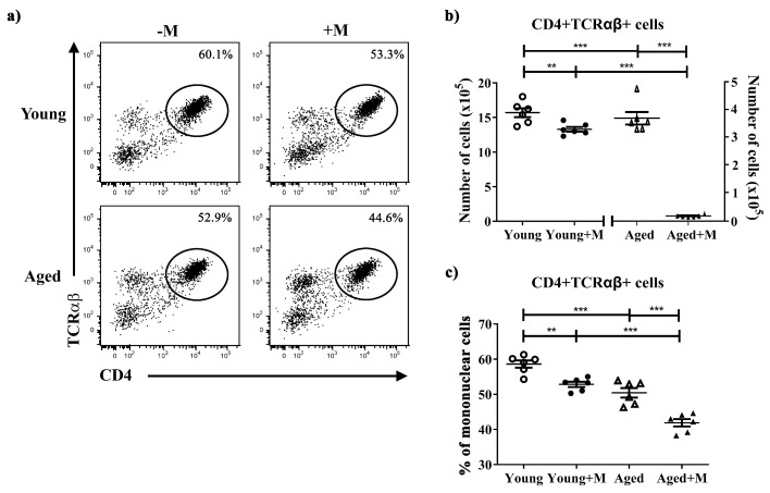
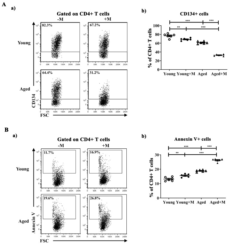
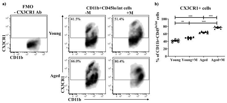
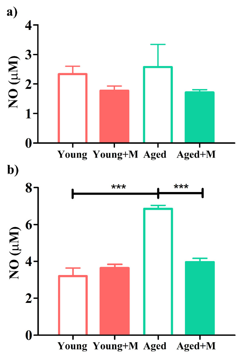
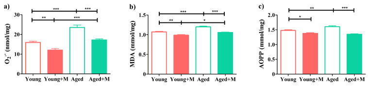
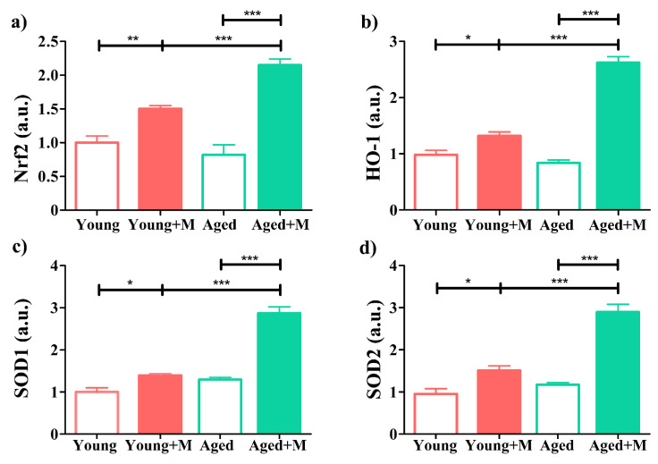
Aging is closely related to the main aspects of multiple sclerosis (MS). The average age of the MS population is increasing and the number of elderly MS patients is expected to increase. In addition to neurons, N-methyl-D-aspartate receptors (NMDARs) are also expressed on non-neuronal cells, such as immune cells. The aim of this study was to investigate the role of NMDARs in experimental autoimmune encephalomyelitis (EAE) in young and aged rats. Memantine, a non-competitive NMDAR antagonist, was administered to young and aged Dark Agouti rats from day 7 after immunization. Antagonizing NMDARs had a more favourable effect on clinical disease, reactivation, and apoptosis of CD4+ T cells in the target organ of aged EAE rats. The expression of the fractalkine receptor CX3CR1 was increased in memantine-treated rats, but to a greater extent in aged rats. Additionally, memantine increased Nrf2 and Nrf2-regulated enzymes' mRNA expression in brain tissue. The concentrations of superoxide anion radicals, malondialdehyde, and advanced oxidation protein products in brain tissue were consistent with previous results. Overall, our results suggest that NMDARs play a more important role in the pathogenesis of EAE in aged than in young rats.

Keywords: EAE; NMDARs; aging; memantine; multiple sclerosis.


Conflict of interest statement

The authors declare no conflicts of interest.

Figures

Figure 1
Figure 1
Memantine treatment reduced the mean neurological score and histological score in rats of both age groups. (Panel (A)) Line graph indicates the mean daily neurological score of EAE in rats treated with memantine (+M) or administered with vehicle (−M) for 7 consecutive days from day 7 to day 13 post-immunization. Data are presented as mean ± SEM (data from 4 experiments, n = 6 rats/group/experiment), with statistical differences determined using a two-way ANOVA; # p < 0.05; ## p < 0.01; ### p < 0.001; # young vs. aged; * p < 0.05; * +M vs. age-matched −M. (Panel (B)) Representative photomicrographs (a) indicate mononuclear cell infiltrations in spinal cord sections of memantine-treated (+M) or administered with vehicle (−M) young and aged rats. The arrows indicate mononuclear cell infiltrates. Bar graph (b) indicates histological scores for spinal cord of memantine-treated and untreated young and aged EAE rats. Histological scoring is described in the Material and Methods section. Data are presented as mean ± SEM (n = 3 rats/group). * p < 0.05; *** p < 0.001.
Figure 2
Figure 2
Memantine reduced the frequency and number of CD4+ T lymphocytes infiltrating the spinal cord of immunized rats of both age groups. (a) Representative flow cytometry dot plots present CD4 vs. TCRαβ staining of mononuclear cells (MNCs) retrieved at the peak of EAE from spinal cords (SCs) of young and aged rats treated with memantine (+M) or vehicle (−M). Numbers in dot plots represent frequency. Scatter plots present (b) the number of CD4+ TCRαβ+ cells and (c) their frequency among MNC cells retrieved from SCs. Data are presented as mean ± SEM from 6 rats per group, with statistical differences determined using a two-way ANOVA. ** p < 0.01; *** p < 0.001.
Figure 3
Figure 3
Memantine reduced reactivation and increased apoptosis of CD4+ T lymphocytes in the SC more effectively in aged than in young EAE rats. Representative flow cytometry dot plots show (a) CD134 (Panel (A)) and (a) Annexin V (Panel (B)) staining of cells gated as CD4+ T lymphocytes among mononuclear cells (MNCs) retrieved at the peak of EAE from spinal cords (SCs) of young and aged rats treated with memantine (+M) or vehicle (−M). Numbers in dot plots represent frequency. Scatter plots present the frequency of (b) CD134+ (Panel (A)) and (b) Annexin V+ (Panel (B)) cells among CD4+ T cells in SCs from young and aged +M and −M EAE rats. Data are presented as mean ± SEM from 6 rats per group. Two-way ANOVA showed significant interaction between the effects of treatment and age for the frequency of CD134+ cells (F(1,20) = 63.22; p < 0.001) and Annexin V+ cells (F(1,20) = 34.34; p < 0.001). ** p < 0.01; *** p < 0.001.
Figure 4
Figure 4
NMDAR antagonist induced lower proportions of IL-17+ and IFN-γ+ cells among CD4+ T cells in SCs of aged rats. Representative flow cytometry dot plots show (a) IL-17 (Panel (A)) and (a) IFN-γ (Panel (B)) staining of cells gated as CD4+ T lymphocytes among mononuclear cells (MNCs) retrieved from spinal cords (SCs) of young and aged rats treated with memantine (+M) or vehicle (−M) at the peak of EAE. Numbers in dot plots represent frequency. Scatter plots present the frequency of (b) IL-17+ (Panel (A)) and (b) IFN-γ+ (Panel (B)) cells among CD4+ T cells in SCs from young and aged +M and −M EAE rats. Data are presented as mean ± SEM from 6 rats per group. Two-way ANOVA showed significant interaction between the effects of treatment and age for the frequency of IL-17+ cells (F(1,20) = 12.88; p < 0.01) and IFN-γ+ cells (F(1,20) = 20.45; p < 0.001). * p < 0.05; *** p < 0.001.
Figure 5
Figure 5
Memantine increased the proportion of CX3CR1-expressing microglia more effectively in aged than in young EAE rats. Representative flow cytometry density plot panels present (a) the frequency of CX3CR1+ cells among CD11b+CD45lo/int microglia retrieved from spinal cord (SC) mononuclear cells (MNCs) at the peak of EAE from young and aged rats treated with memantine (+M) or vehicle (−M). Numbers in dot plots represent frequency. FMO control incubated with secondary antibody (Ab) alone, without anti-CX3CR1 Ab (-CX3CR1 Ab), was used to settle the gating boundary for CX3CR1+ cells. (b) Scatter plots indicate the frequency of CX3CR1+ cells among CD11b+CD45lo/int microglia. Data are presented as mean ± SEM from 6 rats per group. Two-way ANOVA showed significant interaction between the effects of treatment and age for CX3CR1+ microglial cells (F(1,20) = 4.58; p < 0.05). ** p < 0.01; *** p < 0.001.
Figure 6
Figure 6
Memantine reduced the production of nitric oxide by macrophages/microglia obtained from the spinal cord of aged EAE rats. Bar graphs indicate concentration of nitric oxide in overnight mononuclear spinal cord (SC) cell cultures (a) in the medium alone or (b) stimulated with lipopolysaccharide (LPS). Data are presented as mean ± SEM from 6 rats per group. Two-way ANOVA showed significant interaction between the effects of treatment and age for NO concentration determined in LPS-stimulated SC mononuclear cell cultures (F(1,20) = 30.50; p < 0.001). *** p < 0.001.
Figure 7
Figure 7
Memantine was more efficient in reducing oxidative brain tissue damage in aged rats. Bar graphs indicate levels of (a) O2˙, (b) malondialdehyde (MDA), and (c) advanced oxidation protein products (AOPPs) at the peak of EAE in the brain tissue of young and aged EAE rats treated with memantine (+M) or vehicle (−M). Data are presented as mean ± SEM from 6 rats per group. Two-way ANOVA showed significant interaction between the effects of treatment and age for the level of AOPP (F(1,20) = 12.19; p < 0.01). * p < 0.05; ** p < 0.01; *** p < 0.001.
Figure 8
Figure 8
Memantine increased the expression of mRNAs for Nrf2 and Nrf2-regulated enzymes. Bar graphs show the fold change in (a) nuclear factor (erythroid-derived 2)-like 2 (Nrf2), (b) heme oxygenase 1 (HO-1), (c) superoxide dismutase (SOD)1, and (d) SOD2 mRNA expression in brain tissue at the peak of EAE from young and aged rats treated with memantine (+M) or vehicle (−M), as determined by qRT-PCR. Results are normalized to β-actin expression. Data are presented as mean ± SEM from 6 rats per group. Two-way ANOVA showed significant interaction between the effects of treatment and age for Nrf2 (F(1,20) = 127.0; p < 0.001), HO-1 (F(1,20) = 80.06; p < 0.001), SOD1 (F(1,20) = 37.91; p < 0.001), and SOD2 (F(1,20) = 22.45; p < 0.001) mRNA expression. * p < 0.05; ** p < 0.01; *** p < 0.001.

Similar articles

References

    1. Buscarinu M.C., Reniè R., Morena E., Romano C., Bellucci G., Marrone A., Bigi R., Salvetti M., Ristori G. Late-Onset MS: Disease Course and Safety-Efficacy of DMTS. Front. Neurol. 2022;13:829331. doi: 10.3389/fneur.2022.829331. - DOI - PMC - PubMed
    1. Graves J.S., Krysko K.M., Hua L.H., Absinta M., Franklin R.J.M., Segal B.M. Ageing and multiple sclerosis. Lancet Neurol. 2023;22:66–77. doi: 10.1016/S1474-4422(22)00184-3. - DOI - PubMed
    1. Tutuncu M., Tang J., Zeid N.A., Kale N., Crusan D.J., Atkinson E.J., Siva A., Pittock S.J., Pirko I., Keegan B.M., et al. Onset of progressive phase is an age-dependent clinical milestone in multiple sclerosis. Mult. Scler. 2013;19:188–198. doi: 10.1177/1352458512451510. - DOI - PMC - PubMed
    1. Prosperini L., Lucchini M., Ruggieri S., Tortorella C., Haggiag S., Mirabella M., Pozzilli C., Gasperini C. Shift of multiple sclerosis onset towards older age. J. Neurol. Neurosurg. Psychiatry. 2022;93:1137–1139. doi: 10.1136/jnnp-2022-329049. - DOI - PubMed
    1. Solaro C., Ponzio M., Moran E., Tanganelli P., Pizio R., Ribizzi G., Venturi S., Mancardi G.L., Battaglia M.A. The changing face of multiple sclerosis: Prevalence and incidence in an aging population. Mult. Scler. 2015;21:1244–1250. doi: 10.1177/1352458514561904. - DOI - PubMed