

Comparison of MRI Sequences to Predict IDH Mutation Status in Gliomas Using Radiomics-Based Machine Learning
- PMID: 38672080
- PMCID: PMC11048271
- DOI: 10.3390/biomedicines12040725
Comparison of MRI Sequences to Predict IDH Mutation Status in Gliomas Using Radiomics-Based Machine Learning
Abstract
Objectives: Regarding the 2021 World Health Organization (WHO) classification of central nervous system (CNS) tumors, the isocitrate dehydrogenase (IDH) mutation status is one of the most important factors for CNS tumor classification. The aim of our study is to analyze which of the commonly used magnetic resonance imaging (MRI) sequences is best suited to obtain this information non-invasively using radiomics-based machine learning models. We developed machine learning models based on different MRI sequences and determined which of the MRI sequences analyzed yields the highest discriminatory power in predicting the IDH mutation status.
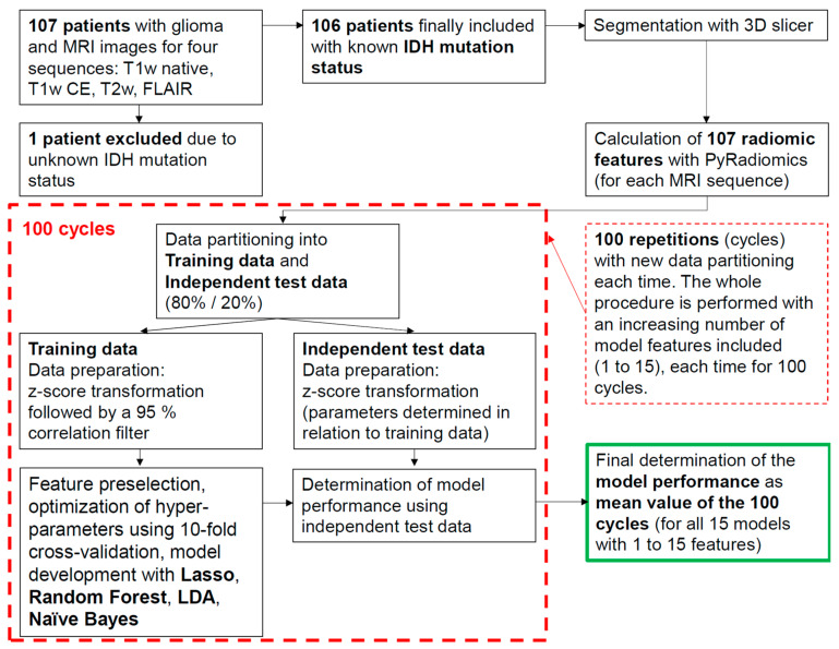
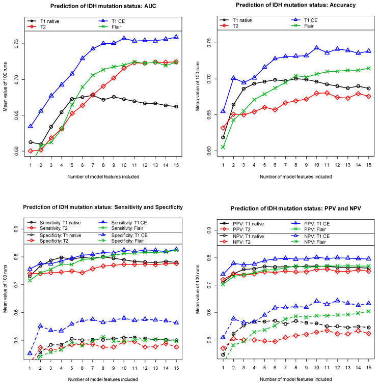
Material and methods: In our retrospective IRB-approved study, we used the MRI images of 106 patients with histologically confirmed gliomas. The MRI images were acquired using the T1 sequence with and without administration of a contrast agent, the T2 sequence, and the Fluid-Attenuated Inversion Recovery (FLAIR) sequence. To objectively compare performance in predicting the IDH mutation status as a function of the MRI sequence used, we included only patients in our study cohort for whom MRI images of all four sequences were available. Seventy-one of the patients had an IDH mutation, and the remaining 35 patients did not have an IDH mutation (IDH wild-type). For each of the four MRI sequences used, 107 radiomic features were extracted from the corresponding MRI images by hand-delineated regions of interest. Data partitioning into training data and independent test data was repeated 100 times to avoid random effects associated with the data partitioning. Feature preselection and subsequent model development were performed using Random Forest, Lasso regression, LDA, and Naïve Bayes. The performance of all models was determined with independent test data.
Results: Among the different approaches we examined, the T1-weighted contrast-enhanced sequence was found to be the most suitable for predicting IDH mutations status using radiomics-based machine learning models. Using contrast-enhanced T1-weighted MRI images, our seven-feature model developed with Lasso regression achieved a mean area under the curve (AUC) of 0.846, a mean accuracy of 0.792, a mean sensitivity of 0.847, and a mean specificity of 0.681. The administration of contrast agents resulted in a significant increase in the achieved discriminatory power.
Conclusions: Our analyses show that for the prediction of the IDH mutation status using radiomics-based machine learning models, among the MRI images acquired with the commonly used MRI sequences, the contrast-enhanced T1-weighted images are the most suitable.
Keywords: IDH mutation status; MRI; artificial intelligence; glioma; machine learning; neuroimaging; radiomics.
Conflict of interest statement
The authors declare no conflicts of interest.
Figures
References
-
- Nicholson J.G., Fine H.A. Diffuse Glioma Heterogeneity and Its Therapeutic Implications. Cancer Discov. 2021;11:575–590. doi: 10.1158/2159-8290.CD-20-1474. - DOI - PubMed
-
- Weller M., van den Bent M., Preusser M., Le Rhun E., Tonn J.C., Minniti G., Bendszus M., Balana C., Chinot O., Dirven L., et al. EANO guidelines on the diagnosis and treatment of diffuse gliomas of adulthood. Nat. Rev. Clin. Oncol. 2021;18:170–186. doi: 10.1038/s41571-020-00447-z. - DOI - PMC - PubMed
-
- WHO Classification of Tumours Editorial Board . Central Nervous System Tumours. 5th ed. Volume 6. International Agency for Research on Cancer; Lyon, France: 2021. [(accessed on 2 October 2023)]. (WHO Classification of Tumours Series). Available online: https://publications.iarc.fr/601.
-
- Cancer Genome Atlas Research Network. Brat D.J., Verhaak R.G., Aldape K.D., Yung W.K., Salama S.R., Cooper L.A., Rheinbay E., Miller C.R., Vitucci M., et al. Comprehensive, Integrative Genomic Analysis of Diffuse Lower-Grade Gliomas. N. Engl. J. Med. 2015;372:2481–2498. doi: 10.1056/NEJMoa1402121. - DOI - PMC - PubMed
LinkOut - more resources
Full Text Sources
