. 2024 Mar 29;12(4):762.
doi: 10.3390/biomedicines12040762.

USP7 Deregulation Impairs S Phase Specific DNA Repair after Irradiation in Breast Cancer Cells

Affiliations

USP7 Deregulation Impairs S Phase Specific DNA Repair after Irradiation in Breast Cancer Cells

Marie Vogt et al. Biomedicines. .

Abstract

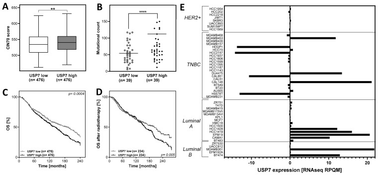
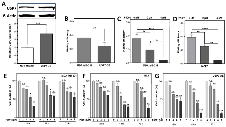
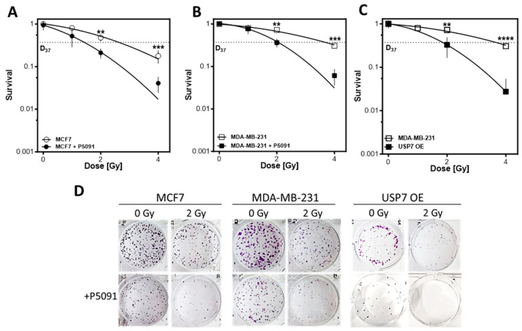
The ubiquitin specific protease 7 (USP7) is a deubiquitinating enzyme with numerous substrates. Aberrant expression of USP7 is associated with tumor progression. This study aims to investigate how a deregulated USP7 expression affects chromosomal instability and prognosis of breast cancer patients in silico and radiosensitivity and DNA repair in breast cancer cells in vitro. The investigations in silico were performed using overall survival and USP7 mRNA expression data of breast cancer patients. The results showed that a high USP7 expression was associated with increased chromosomal instability and decreased overall survival. The in vitro experiments were performed in a luminal and a triple-negative breast cancer cell line. Proliferation, DNA repair, DNA replication stress, and survival after USP7 overexpression or inhibition and irradiation were analyzed. Both, USP7 inhibition and overexpression resulted in decreased cellular survival, distinct radiosensitization and an increased number of residual DNA double-strand breaks in the S phase following irradiation. RAD51 recruitment and base incorporation were decreased after USP7 inhibition plus irradiation and more single-stranded DNA was detected. The results show that deregulation of USP7 activity disrupts DNA repair in the S phase by increasing DNA replication stress and presents USP7 as a promising target to overcome the radioresistance of breast tumors.

Keywords: DNA repair; USP7; breast cancer; genomic instability; radiosensitization.


Conflict of interest statement

The authors declare no conflicts of interest.

Figures

Figure 1
Figure 1
High expression of USP7 correlates with chromosomal instability and survival in breast cancer patients. (A): CIN70 score in tumors, sorted by USP7 mRNA expression. Plotted are the extreme quartiles (n = 952 out of n = 1904). (B): Mutational count in patients, sorted by USP7 mRNA expression. Plotted are the extreme quartiles (n = 78 out of n = 156). (C): Kaplan-Meier analysis of USP7 mRNA expression as prognostic factor for overall survival (OS) in breast cancer patients, using the extreme quartiles and analysis with the log-rank test (n = 952 out of n = 1904) or (D): Only breast cancer patients who received radiotherapy (n = 568 out of n = 1136). (E): USP7 mRNA expression in breast cancer cell lines, sorted by molecular subtype. Clinical- and mRNA expression data were extracted from the METABRIC dataset, provided by the cBioportal database (http://www.cbioportal.org (accessed on 14 June 2023)). The CIN70 score was calculated according to Birkbak et al. [37] by adding the expression values of all CIN70 genes from n = 1904 patients. For the mutational count, clinical and mRNA expression data were extracted and the extreme quartiles of the number of mutations per tumor were plotted. The USP7 mRNA expression data in cell lines were extracted from the Cancer Cell Line Encyclopedia dataset and sorted by molecular subtype. Asterisks represent significant differences (** p < 0.01, **** p < 0.0001, Student’s t-test).
Figure 2
Figure 2
Both, up- and downregulation of USP7 reduces cellular proliferation in breast cancer cells. (A): Immunodetection of USP7. MDA-MB-231 cells were stably transfected with pQFlag-USP7 WT plasmid, total protein was extracted from pooled clones. Separated proteins were transferred and detected by appropriate antibodies, with ß-Actin serving as loading control. (BD): Plating efficiency of USP7 overexpressing (USP7 OE) or with P5091 inhibited cells. Colonies were stained and counted. (EG): Proliferation of cells treated with P5091. Proliferating cells were seeded, treated with indicated doses of P5091 and the cell number was quantified as indicated. Shown are means of three independent experiments ± SEM (n.s. not significant; * p < 0.05; ** p < 0.01, *** p < 0.001, **** p < 0.0001, Student’s t-test).
Figure 3
Figure 3
USP7 inhibition and overexpression lead to an increase in DNA damage in the S phase. (A): Exemplary depiction of 53BP1 foci 24 h after 6 Gy compared to control. (B): Quantification of 53BP1 foci 4 h or (C): 24 h after irradiation in the presence or absence of USP7 inhibition. (D): Exemplary depiction of yH2AX and PCNA foci 4 h after irradiation or control. (E): yH2AX foci in PCNA positive cells or (F): PCNA negative cells after irradiation with and without USP7 inhibition. Cells were treated with 4 µm of P5091, irradiated with 6 Gy and immunofluorescence staining was performed 4 h or 24 h after treatment with 53BP1 or 6 h after treatment with PCNA and yH2AX and fluorescent secondary antibodies. Foci were quantified automatically by the Medipan AKLIDES® device. Shown are the means of three independent experiments ± SEM. Asterisks represent significant differences (n.s. not significant; * p < 0.05, *** p < 0.001, **** p < 0.0001, Student’s t-test).
Figure 4
Figure 4
USP7 inhibition and overexpression leads to increased DNA replication stress. (A): Examples of RAD51 foci or (C): RPA foci or (E): PCNA foci after 4/6 h with or without irradiation. (B): Quantification of RAD51 foci in RAD51 positive cells, 6 h or (D): of RPA-foci 4 h or (F): of PCNA foci 4 h after irradiation. Cells were treated with 4 µM P5091 and irradiated with 6 Gy. Fixation and immunofluorescence staining were performed 4/6 h after irradiation with the indicated antibodies plus nuclear staining using DAPI. Foci were quantified automatically by the Medipan AKLIDES® device. (G): Exemplary depiction of EdU positive cells after irradiation, with or without USP7 inhibition. (H): EdU incorporation after irradiation. Cells were pulse-labeled with EdU and treated with either- or both P5091 and 6 Gy of ionizing radiation. Incorporated EdU was stained and EdU positive cells were captured. Fluorescence intensity was analyzed with the Image J software. Means are depicted in red. Shown are the means of three independent experiments ± SEM. Asterisks represent significant differences (n.s. not significant; ** p < 0.01, *** p < 0.001, **** p < 0.0001, Student’s t-test).
Figure 5
Figure 5
USP7 inhibition and overexpression radiosensitize breast cancer cell lines independent of their p53 status and molecular subtype. (AC): Cellular survival after treatment with irradiation alone or in combination with the USP7 inhibitor P5091. Cells were plated, treated with P5091 (2 µM) and/or irradiation, fixed and stained after 14 days and the number of colonies was counted. Shown are the means of three independent experiments ± SEM. Asterisks represent significant differences (** p < 0.01, *** p < 0.001, **** p < 0.0001, Student’s t-test). (D): Exemplary pictures from colony assays of MCF7, MDA-MB-231 and USP7 OE cells after 2 Gy irradiation, with or without P5091.

Similar articles

Cited by

References

    1. Sung H., Ferlay J., Siegel R.L., Laversanne M., Soerjomataram I., Jemal A., Bray F. Global Cancer Statistics 2020: GLOBOCAN Estimates of Incidence and Mortality Worldwide for 36 Cancers in 185 Countries. CA Cancer J. Clin. 2021;71:209–249. doi: 10.3322/caac.21660. - DOI - PubMed
    1. Prat A., Pineda E., Adamo B., Galván P., Fernández A., Gaba L., Díez M., Viladot M., Arance A., Muñoz M. Clinical implications of the intrinsic molecular subtypes of breast cancer. Breast. 2015;24((Suppl. 2)):S26–S35. doi: 10.1016/j.breast.2015.07.008. - DOI - PubMed
    1. Ades F., Zardavas D., Bozovic-Spasojevic I., Pugliano L., Fumagalli D., de Azambuja E., Viale G., Sotiriou C., Piccart M. Luminal B breast cancer: Molecular characterization, clinical management, and future perspectives. J. Clin. Oncol. 2014;32:2794–2803. doi: 10.1200/JCO.2013.54.1870. - DOI - PubMed
    1. O’Brien K.M., Cole S.R., Tse C.-K., Perou C.M., Carey L.A., Foulkes W.D., Dressler L.G., Geradts J., Millikan R.C. Intrinsic breast tumor subtypes, race, and long-term survival in the Carolina Breast Cancer Study. Clin. Cancer Res. 2010;16:6100–6110. doi: 10.1158/1078-0432.CCR-10-1533. - DOI - PMC - PubMed
    1. Carey L.A., Perou C.M., Livasy C.A., Dressler L.G., Cowan D., Conway K., Karaca G., Troester M.A., Tse C.K., Edmiston S., et al. Race, breast cancer subtypes, and survival in the Carolina Breast Cancer Study. JAMA. 2006;295:2492–2502. doi: 10.1001/jama.295.21.2492. - DOI - PubMed