

Using Macrophage Polarization in Human Platelet Lysate to Test the Immunomodulatory Potential of Cells for Clinical Use
- PMID: 38672188
- PMCID: PMC11048141
- DOI: 10.3390/biomedicines12040833
Using Macrophage Polarization in Human Platelet Lysate to Test the Immunomodulatory Potential of Cells for Clinical Use
Abstract
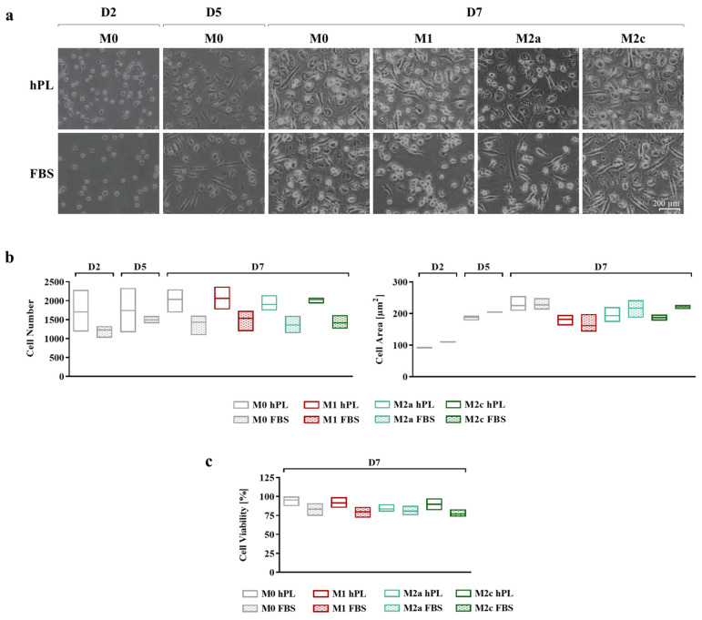
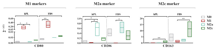
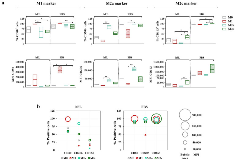
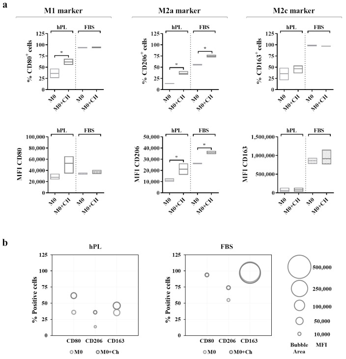
Macrophage-based co-cultures are used to test the immunomodulatory function of candidate cells for clinical use. This study aimed to characterize a macrophage polarization model using human platelet lysate (hPL) as a GMP-compliant alternative to Fetal Bovine Serum (FBS). Primary human monocytes were differentiated into unpolarized (M0) or polarized (M1, M2a, and M2c) macrophages in an hPL- or FBS-based medium. The protein secretion profiles and expression of phenotypic markers (CD80 for M1, CD206 for M2a, and CD163 for M2c) were analyzed. Subsequently, chondrocytes were tested in an hPL-based co-culture model to assess their immunomodulatory function in view of their possible use in patients with osteoarthritis. The results showed similar marker regulation between hPL and FBS cultures, but lower basal levels of CD206 and CD163 in hPL-cultured macrophages. Functional co-culture experiments with chondrocytes revealed increased CD206 expression both in hPL and in FBS, indicating an interaction between macrophages and chondrocytes. While markers in FBS-cultured macrophages were confirmed in hPL-cultured cells, the interpretation of marker modulation in immunomodulatory assays with hPL-based cultures should be carried out cautiously due to the observed differences in the basal marker levels for CD206 and CD163. This research underscores the utility of hPL as a GMP-compliant alternative to FBS for macrophage-based co-cultures and highlights the importance of understanding marker expressions in different culture conditions.
Keywords: chondrocyte; co-culture; human platelet lysate; macrophage; polarization.
Conflict of interest statement
The authors declare no conflicts of interest. The funders had no role in the design of the study; in the collection, analyses, or interpretation of data; in the writing of the manuscript; or in the decision to publish the results.
Figures


Similar articles
-
Jaw Periosteum-Derived Mesenchymal Stem Cells Regulate THP-1-Derived Macrophage Polarization.Int J Mol Sci. 2021 Apr 21;22(9):4310. doi: 10.3390/ijms22094310. Int J Mol Sci. 2021. PMID: 33919221 Free PMC article.
-
Feasibility of Human Platelet Lysate as an Alternative to Foetal Bovine Serum for In Vitro Expansion of Chondrocytes.Int J Mol Sci. 2021 Jan 28;22(3):1269. doi: 10.3390/ijms22031269. Int J Mol Sci. 2021. PMID: 33525349 Free PMC article. Review.
-
Human Platelet Lysate as Alternative of Fetal Bovine Serum for Enhanced Human In Vitro Bone Resorption and Remodeling.Front Immunol. 2022 Jun 20;13:915277. doi: 10.3389/fimmu.2022.915277. eCollection 2022. Front Immunol. 2022. PMID: 35795685 Free PMC article.
-
The Upscale Manufacture of Chondrocytes for Allogeneic Cartilage Therapies.Tissue Eng Part C Methods. 2023 Sep;29(9):424-437. doi: 10.1089/ten.TEC.2023.0037. Tissue Eng Part C Methods. 2023. PMID: 37395490 Free PMC article.
-
Systematic review and meta-analysis on the use of human platelet lysate for mesenchymal stem cell cultures: comparison with fetal bovine serum and considerations on the production protocol.Stem Cell Res Ther. 2022 Apr 4;13(1):142. doi: 10.1186/s13287-022-02815-1. Stem Cell Res Ther. 2022. PMID: 35379348 Free PMC article.
References
Grants and funding
LinkOut - more resources
Full Text Sources
Research Materials
