. 2024 Apr 12;12(4):858.
doi: 10.3390/biomedicines12040858.

Significant Genes Associated with Mortality and Disease Progression in Grade II and III Glioma

Affiliations

Significant Genes Associated with Mortality and Disease Progression in Grade II and III Glioma

Bo Mi Choi et al. Biomedicines. .

Abstract

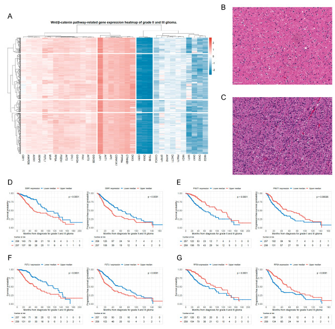
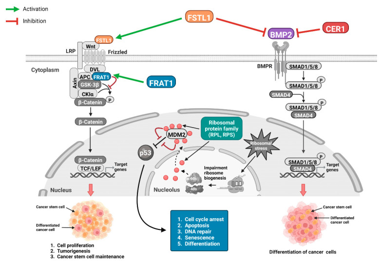
Background: The Wnt/β-catenin pathway plays a critical role in the tumorigenesis and maintenance of glioma stem cells. This study aimed to evaluate significant genes associated with the Wnt/β-catenin pathway involved in mortality and disease progression in patients with grade II and III glioma, using the Cancer Genome Atlas (TCGA) database.

Methods: We obtained clinicopathological information and mRNA expression data from 515 patients with grade II and III gliomas from the TCGA database. We performed a multivariate Cox regression analysis to identify genes independently associated with glioma prognosis.

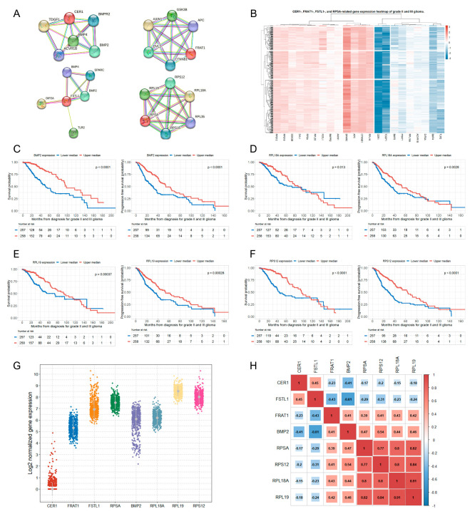
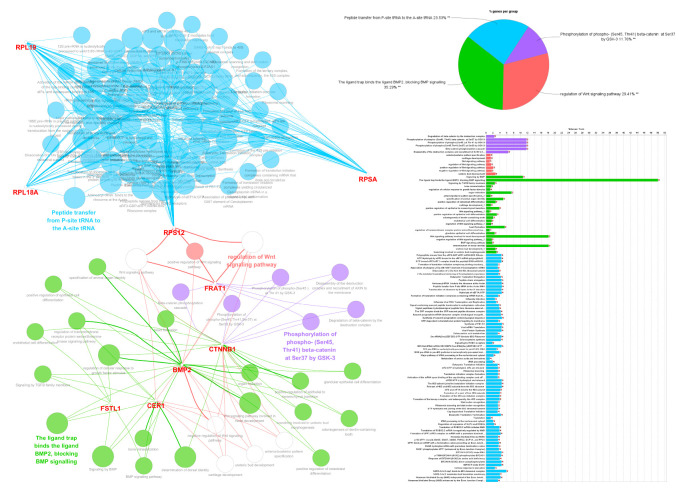
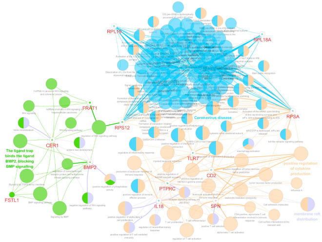
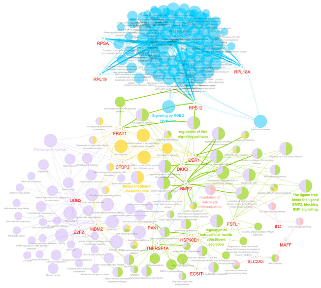
Results: The analysis of 34 genes involved in Wnt/β-catenin signaling demonstrated that four genes (CER1, FRAT1, FSTL1, and RPSA) related to the Wnt/β-catenin pathway were significantly associated with mortality and disease progression in patients with grade II and III glioma. We also identified additional genes related to the four significant genes of the Wnt/β-catenin pathway mentioned above. The higher expression of BMP2, RPL18A, RPL19, and RPS12 is associated with better outcomes in patients with glioma.

Conclusions: Using a large-scale open database, we identified significant genes related to the Wnt/β-catenin signaling pathway associated with mortality and disease progression in patients with grade II and III gliomas.

Keywords: TCGA; Wnt/β-catenin signaling; gene; glioma; survival.


Conflict of interest statement

The authors declare no conflicts of interest.

Figures

Figure 1
Figure 1
Wnt/β-catenin pathway-related gene expression pattern in patients with grade II and III glioma. Four significant genes related to the Wnt/β-catenin pathway are associated with mortality and disease progression in patients with grade II and III glioma. (A) A hierarchically clustered heatmap showing the expression patterns of 34 genes related to the Wnt/β-catenin signaling pathway in patients with grade II and III glioma. Gene expressions are transformed in log2, and color density is displayed, indicating levels of log2 fold changes. Red and blue represent up- and down-regulated expressions in grade II and III glioma, respectively; (B) CNS WHO-grade 2 glioma: an infiltrating astrocytoma of low cell density, showing mild nuclear atypia of tumor cells and a dense fibrillar background with mild edema; (C) CNS WHO-grade 3 glioma: IDH-mutant astrocytoma showing greater cellularity, nuclear atypia, and increased mitotic activity than that exhibited by WHO-grade 2 astrocytoma; (D) OS and PFS rates of patients with glioma based on the upper and lower median groups of CER1 expression; (E) OS and PFS rates of patients with glioma based on the upper and lower median groups of FRAT1 expression; (F) OS and PFS rates of patients with glioma based on the upper and lower median groups of FSTL1 expression; and (G) OS and PFS rates of patients with glioma based on the upper and lower median groups of RPSA expression. CNS, central nervous system; WHO, World Health Organization; IDH, isocitrate dehydrogenase; OS, overall survival; PFS, progression-free survival; CER1, cerebellar 1; FRAT1, FRAT regulator of WNT signaling pathway 1; FSTL1, follistatin-like 1; RPSA, ribosomal protein SA.
Figure 2
Figure 2
Identification of additional four significant genes associated with mortality and disease progression in patients with grade II and III glioma and correlations between eight significant genes. (A) The Wnt/β-catenin pathway-associated protein–protein interaction network of four significant genes (CER1, FRAT1, FSTL1, and RPSA) was constructed using a STRING database (V11.5). Five related genes were searched for each significant gene. The thickness of the line between any two proteins represents the degree of confidence in the interaction between the two proteins, with thicker lines indicating higher confidence; (B) a hierarchically clustered heatmap showing the expression patterns of the expressions of eighteen genes related to the four significant genes (CER1, FRAT1, FSTL1, and RPSA) in patients with grade II and III glioma. Gene expressions are transformed in log2, and color density is displayed, indicating log2 fold changes. Red and blue represent up- and downregulated expressions in grade II and III glioma, respectively; OS and PFS rates of patients with glioma according to the upper and lower median groups of (C) BMP2 expression; (D) RPL18A expression; (E) RPL19 expression; and (F) RPS12 expression; (G) strip plots showing log2-transformed gene mRNA expressions based on the selected eight significant genes; and (H) Pearson’s correlation coefficients and significance levels were calculated between the selected eight significant genes. The color-coordinated legend indicates the value and sign of Pearson’s correlation coefficient. The number in the box indicates Pearson’s correlation coefficient. The x in the box indicates a p-value of ≥0.001. CER1, cerebrum 1; FRAT1, FRAT regulator of WNT signaling pathway 1; FSTL1, follistatin-like 1; RPSA, ribosomal protein SA; STRING, Search Tool for the Retrieval of Interacting Genes/Proteins; OS, overall survival; PFS, progression-free survival; BMP2, bone morphogenetic protein 2; RPL18A, ribosomal protein L18A; RPL19, ribosomal protein L19; RPS12, ribosomal protein S12.
Figure 3
Figure 3
Bioinformatics analysis using Cytoscape with ClueGo and CluePedia plug-ins. The grouping of the networks of significant genes associated with the prognosis of grade II and III gliomas based on functionally enriched GO terms and pathways. GO, gene ontology.
Figure 4
Figure 4
Bioinformatics network analysis visualizing the interactions between eight significant genes identified in this study (CER1, FRAT1, FSTL1, RPSA, BMP2, RPL18A, RPL19, and RPS12) and genes that are potentially involved in regulating the immune microenvironment and serve as independent prognostic markers for LGG (CD2, SPN, IL18, PTPRC, GZMA, and TLR7). The network was generated using Cytoscape with functional GO terms and biological pathways enrichment. CER1, cerberus 1; FRAT1, FRAT regulator of WNT signaling pathway 1; FSTL1, follistatin-like 1; RPSA, ribosomal protein SA; BMP2, bone morphogenetic protein 2; RPL18A, ribosomal protein L18A; RPL19, ribosomal protein L19; RPS12, ribosomal protein S12; LGG, lower-grade glioma; SPN, sialophorin; IL, interleukin; PTPRC; protein tyrosine phosphatase receptor type C; GZMA, granzyme A; TLR7, Toll-like receptor 7; GO, gene ontology.
Figure 5
Figure 5
Bioinformatics network analysis visualizing the interactions between eight significant genes discovered in this research (CER1, FRAT1, FSTL1, RPSA, BMP2, RPL18A, RPL19, and RPS12), and twelve independent genes from ten oncogenic signaling pathways (E2F2 [cell cycle signaling pathway], CTBP2 [Notch signaling], MAFF [Nrf2 signaling], SLC2A3 [Nrf2 signaling], ECSIT [PI3K signaling], HSP90B1 [PI3K signaling], TNFRSF1A [PI3K signaling], PAK1 [RTK signaling], ID4 [TGF-β signaling], DDB2 [p53 signaling], MDM2 [p53 and cell cycle signaling], and DKK3 [Wnt/β-catenin signaling]) that have been significantly associated with prognosis in patients with GBM. The network was generated using Cytoscape with the functional enrichment of GO terms and biological pathways. CER1, cerberus 1; FRAT1, FRAT regulator of WNT signaling pathway 1; FSTL1, follistatin like 1; RPSA, ribosomal protein SA; BMP2, bone morphogenetic protein 2; RPL18A, ribosomal protein L18A; RPL19, ribosomal protein L19; RPS12, ribosomal protein S12; E2F2, E2F transcription factor 2; CTBP2, C-terminal-binding protein 2; MAFF, MAF bZIP transcription factor F; Nrf2, nuclear factor erythroid 2-related factor 2; SLC2A3, solute carrier family 2 member 3; ECSIT, evolutionarily conserved signaling intermediate in Toll pathways; PI3K, phosphatidylinositol 3-kinase; HSP90B1, heat shock protein 90 kDa beta member 1; TNFRSF1A, tumor necrosis factor receptor superfamily member 1A; PAK1, p21 activated kinase 1; RTK, receptor tyrosine kinase; ID4, inhibitor of DNA binding 4; TGF-β, transforming growth factor beta; DDB2, damage-specific DNA-binding protein 2; MDM2, mouse double minute 2 homolog; DKK3, dickkopf-3; GBM, glioblastoma multiforme; GO, gene ontology.
Figure 6
Figure 6
Schematic illustrations of possible roles of the eight significant genes in glioma: Overexpression of FSTL1 activates the Wnt/β-catenin pathway to induce tumorigenesis and cancer stem cell maintenance and inhibits the BMP2 pathway, which leads to the undifferentiation of cancer cells. Increased CER1 expression suppresses BMP2 to induce undifferentiated glioma cells. FRAT1 is known to act on the GSK3 signaling network of the Wnt/β-catenin signaling to activate β-catenin. However, based on our findings, the function of FRAT1 may not be limited to Wnt signaling. It can perform other functions independently of its role in the GSK3 signaling network of Wnt signaling. The overexpression of specific RPs suppresses the p53 inhibition of MDM2, which may lead to glioma cell cycle arrest, apoptosis, and the differentiation of glioma cells. FSTL1, follistatin-like 1; BMP2, bone morphogenetic protein 2; CER1, cerebellar 1; FRAT1, FRAT regulator of WNT signaling pathway 1; GSK3, glycogen synthase kinase 3; RP, ribosomal protein; MDM, mouse double minute 2 homolog.

Similar articles

Cited by

References

    1. Mesfin F.B., Al-Dhahir M.A. StatPearls. StatPearls Publishing; Treasure Island, FL, USA: 2022. Gliomas.
    1. Miranda A., Hamilton P.T., Zhang A.W., Pattnaik S., Becht E., Mezheyeuski A., Bruun J., Micke P., de Reynies A., Nelson B.H. Cancer Stemness, Intratumoral Heterogeneity, and Immune Response across Cancers. Proc. Natl. Acad. Sci. USA. 2019;116:9020–9029. doi: 10.1073/pnas.1818210116. - DOI - PMC - PubMed
    1. Fodde R., Brabletz T. Wnt/Beta-Catenin Signaling in Cancer Stemness and Malignant Behavior. Curr. Opin. Cell Biol. 2007;19:150–158. doi: 10.1016/j.ceb.2007.02.007. - DOI - PubMed
    1. Chen Y.-H., McGowan L.D., Cimino P.J., Dahiya S., Leonard J.R., Lee D.Y., Gutmann D.H. Mouse Low-Grade Gliomas Contain Cancer Stem Cells with Unique Molecular and Functional Properties. Cell Rep. 2015;10:1899–1912. doi: 10.1016/j.celrep.2015.02.041. - DOI - PMC - PubMed
    1. Denysenko T., Annovazzi L., Cassoni P., Melcarne A., Mellai M., Schiffer D. WNT/β-Catenin Signaling Pathway and Downstream Modulators in Low- and High-Grade Glioma. Cancer Genom. Proteom. 2016;13:31–45. - PubMed

LinkOut - more resources