

Understanding Tendon Fibroblast Biology and Heterogeneity
- PMID: 38672213
- PMCID: PMC11048404
- DOI: 10.3390/biomedicines12040859
Understanding Tendon Fibroblast Biology and Heterogeneity
Abstract
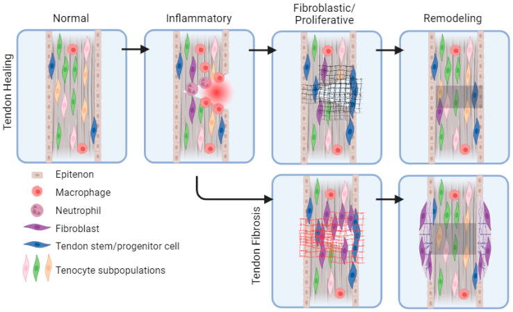
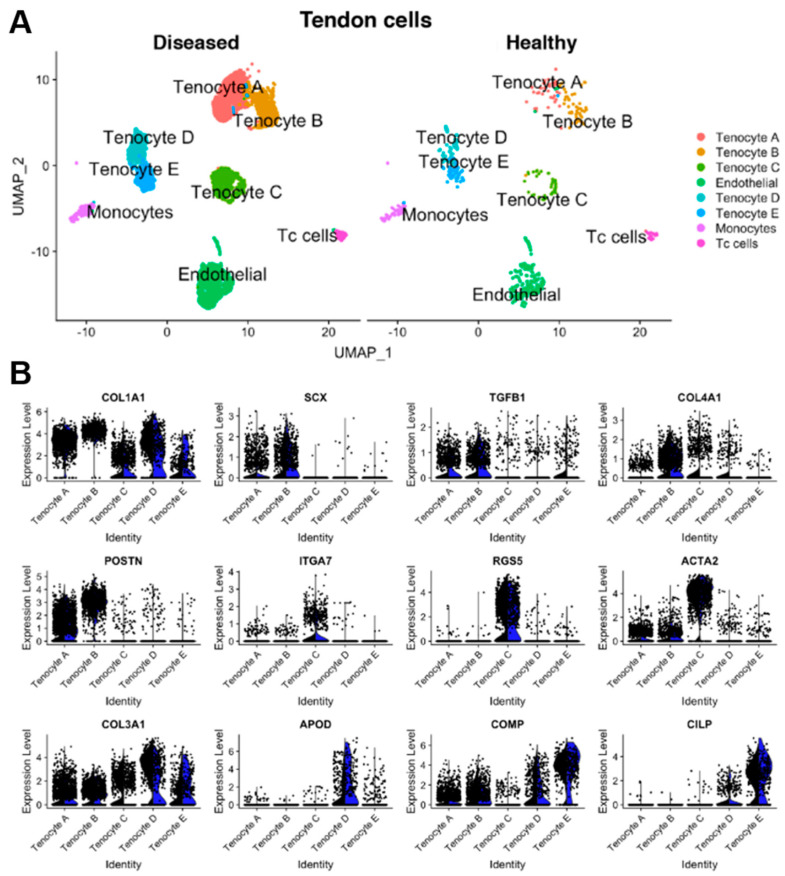
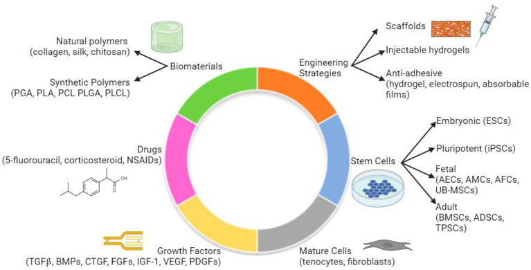
Tendon regeneration has emerged as an area of interest due to the challenging healing process of avascular tendon tissue. During tendon healing after injury, the formation of a fibrous scar can limit tendon strength and lead to subsequent complications. The specific biological mechanisms that cause fibrosis across different cellular subtypes within the tendon and across different tendons in the body continue to remain unknown. Herein, we review the current understanding of tendon healing, fibrosis mechanisms, and future directions for treatments. We summarize recent research on the role of fibroblasts throughout tendon healing and describe the functional and cellular heterogeneity of fibroblasts and tendons. The review notes gaps in tendon fibrosis research, with a focus on characterizing distinct fibroblast subpopulations in the tendon. We highlight new techniques in the field that can be used to enhance our understanding of complex tendon pathologies such as fibrosis. Finally, we explore bioengineering tools for tendon regeneration and discuss future areas for innovation. Exploring the heterogeneity of tendon fibroblasts on the cellular level can inform therapeutic strategies for addressing tendon fibrosis and ultimately reduce its clinical burden.
Keywords: biomaterials; fibroblast; fibrosis; tendon; tenocyte.
Conflict of interest statement
The authors declare no conflicts of interest.
Figures
Similar articles
-
The cellular basis of fibrotic tendon healing: challenges and opportunities.Transl Res. 2019 Jul;209:156-168. doi: 10.1016/j.trsl.2019.02.002. Epub 2019 Feb 8. Transl Res. 2019. PMID: 30776336 Free PMC article. Review.
-
Biomaterials strategies to balance inflammation and tenogenesis for tendon repair.Acta Biomater. 2021 Aug;130:1-16. doi: 10.1016/j.actbio.2021.05.043. Epub 2021 May 31. Acta Biomater. 2021. PMID: 34082095 Review.
-
Scleraxis lineage cells contribute to organized bridging tissue during tendon healing and identify a subpopulation of resident tendon cells.FASEB J. 2019 Jul;33(7):8578-8587. doi: 10.1096/fj.201900130RR. Epub 2019 Apr 5. FASEB J. 2019. PMID: 30951381 Free PMC article.
-
The mechanisms and functions of TGF-β1 in tendon healing.Injury. 2023 Nov;54(11):111052. doi: 10.1016/j.injury.2023.111052. Epub 2023 Sep 15. Injury. 2023. PMID: 37738787 Review.
-
Repair of tendon defect with dermal fibroblast engineered tendon in a porcine model.Tissue Eng. 2006 Apr;12(4):775-88. doi: 10.1089/ten.2006.12.775. Tissue Eng. 2006. PMID: 16674291
Cited by
-
Impact of Composition and Autoclave Sterilization on the Mechanical and Biological Properties of ECM-Mimicking Cryogels.Polymers (Basel). 2024 Jul 7;16(13):1939. doi: 10.3390/polym16131939. Polymers (Basel). 2024. PMID: 39000793 Free PMC article.
-
Mesenchymal stem cell activity across a graded scaffold-hydrogel composite biomaterial for tendon-to-bone enthesis repair.Bioact Mater. 2025 Jul 15;53:287-299. doi: 10.1016/j.bioactmat.2025.07.017. eCollection 2025 Nov. Bioact Mater. 2025. PMID: 40703334 Free PMC article.
-
[A comparative study of dynamic versus static rehabilitation protocols after acute Achilles tendon rupture repair with channel assisted minimally invasive repair technique].Zhongguo Xiu Fu Chong Jian Wai Ke Za Zhi. 2024 Dec 15;38(12):1492-1498. doi: 10.7507/1002-1892.202408024. Zhongguo Xiu Fu Chong Jian Wai Ke Za Zhi. 2024. PMID: 39694840 Free PMC article. Chinese.
-
Diseased Tendon Models Demonstrate Influence of Extracellular Matrix Alterations on Extracellular Vesicle Profile.Bioengineering (Basel). 2024 Oct 12;11(10):1019. doi: 10.3390/bioengineering11101019. Bioengineering (Basel). 2024. PMID: 39451395 Free PMC article.
References
-
- Miller K., Hsu J.E., Soslowsky L.J. Materials in Tendon and Ligament Repair. Compr. Biomater. 2011;6:257–279. doi: 10.1016/B978-0-08-055294-1.00218-X. - DOI
Publication types
LinkOut - more resources
Full Text Sources
