

Advancing Surgical Arrhythmia Ablation: Novel Insights on 3D Printing Applications and Two Biocompatible Materials
- PMID: 38672223
- PMCID: PMC11048352
- DOI: 10.3390/biomedicines12040869
Advancing Surgical Arrhythmia Ablation: Novel Insights on 3D Printing Applications and Two Biocompatible Materials
Abstract
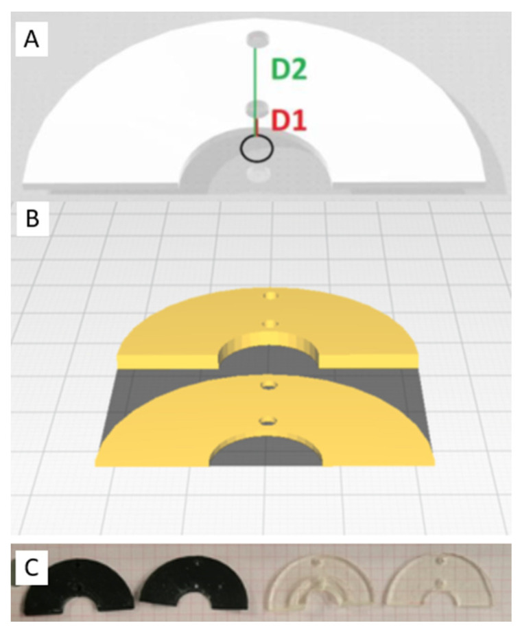
To date, studies assessing the safety profile of 3D printing materials for application in cardiac ablation are sparse. Our aim is to evaluate the safety and feasibility of two biocompatible 3D printing materials, investigating their potential use for intra-procedural guides to navigate surgical cardiac arrhythmia ablation. Herein, we 3D printed various prototypes in varying thicknesses (0.8 mm-3 mm) using a resin (MED625FLX) and a thermoplastic polyurethane elastomer (TPU95A). Geometrical testing was performed to assess the material properties pre- and post-sterilization. Furthermore, we investigated the thermal propagation behavior beneath the 3D printing materials during cryo-energy and radiofrequency ablation using an in vitro wet-lab setup. Moreover, electron microscopy and Raman spectroscopy were performed on biological tissue that had been exposed to the 3D printing materials to assess microparticle release. Post-sterilization assessments revealed that MED625FLX at thicknesses of 1 mm, 2.5 mm, and 3 mm, along with TPU95A at 1 mm and 2.5 mm, maintained geometrical integrity. Thermal analysis revealed that material type, energy source, and their factorial combination with distance from the energy source significantly influenced the temperatures beneath the 3D-printed material. Electron microscopy revealed traces of nitrogen and sulfur underneath the MED625FLX prints (1 mm, 2.5 mm) after cryo-ablation exposure. The other samples were uncontaminated. While Raman spectroscopy did not detect material release, further research is warranted to better understand these findings for application in clinical settings.
Keywords: ablation; additive manufacturing; cardiac; epicardial; material testing; three-dimensional printing (3D printing).
Conflict of interest statement
M.L.M. is a consultant for AtriCure. C.d.A. receives research grants and compensation for teaching purposes and proctoring from AtriCure. R.K. received a one-time consultant fee from AtriCure. The other authors declare no conflicts of interest.
Figures


Similar articles
-
3D-Printed Biomaterial Testing in Response to Cryoablation: Implications for Surgical Ventricular Tachycardia Ablation.J Clin Med. 2023 Jan 29;12(3):1036. doi: 10.3390/jcm12031036. J Clin Med. 2023. PMID: 36769681 Free PMC article.
-
Temperature analysis of 3D-printed biomaterials during unipolar and bipolar radiofrequency ablation procedure.Front Cardiovasc Med. 2022 Sep 14;9:978333. doi: 10.3389/fcvm.2022.978333. eCollection 2022. Front Cardiovasc Med. 2022. PMID: 36186978 Free PMC article.
-
Development of a 3D printed surgical guide for Brugada syndrome substrate ablation.Front Cardiovasc Med. 2022 Nov 15;9:1029685. doi: 10.3389/fcvm.2022.1029685. eCollection 2022. Front Cardiovasc Med. 2022. PMID: 36457802 Free PMC article.
-
How useful is 3D printing in maxillofacial surgery?J Stomatol Oral Maxillofac Surg. 2017 Sep;118(4):206-212. doi: 10.1016/j.jormas.2017.07.002. Epub 2017 Jul 18. J Stomatol Oral Maxillofac Surg. 2017. PMID: 28732777 Review.
-
Polymer 3D Printing Review: Materials, Process, and Design Strategies for Medical Applications.Polymers (Basel). 2021 May 6;13(9):1499. doi: 10.3390/polym13091499. Polymers (Basel). 2021. PMID: 34066639 Free PMC article. Review.
Cited by
-
Research on the preparation of antimicrobial material based on thermoplastic polyurethane and drug release control.Drug Deliv Transl Res. 2025 Jul;15(7):2534-2546. doi: 10.1007/s13346-024-01751-2. Epub 2024 Dec 2. Drug Deliv Transl Res. 2025. PMID: 39623244
References
-
- Olivieri L.J., Su L., Hynes C.F., Krieger A., Alfares F.A., Ramakrishnan K., Zurakowski D., Marshall M.B., Kim P.C.W., Jonas R.A., et al. “Just-In-Time” Simulation Training Using 3-D Printed Cardiac Models after Congenital Cardiac Surgery. World J. Pediatr. Congenit. Heart Surg. 2016;7:164–168. doi: 10.1177/2150135115623961. - DOI - PubMed
-
- Talevi G., Pannone L., Monaco C., Bori E., Cappello I.A., Candelari M., Ramak R., La Meir M., Gharaviri A., Chierchia G.B., et al. Development of a 3D Printed Surgical Guide for Brugada Syndrome Substrate Ablation. Front. Cardiovasc. Med. 2022;9:1029685. doi: 10.3389/fcvm.2022.1029685. - DOI - PMC - PubMed
LinkOut - more resources
Full Text Sources
Miscellaneous
