. 2024 Apr 18;12(4):904.
doi: 10.3390/biomedicines12040904.

Verapamil Attenuates the Severity of Tendinopathy by Mitigating Mitochondrial Dysfunction through the Activation of the Nrf2/HO-1 Pathway

Affiliations

Verapamil Attenuates the Severity of Tendinopathy by Mitigating Mitochondrial Dysfunction through the Activation of the Nrf2/HO-1 Pathway

Zengguang Wang et al. Biomedicines. .

Abstract

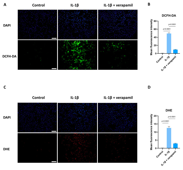
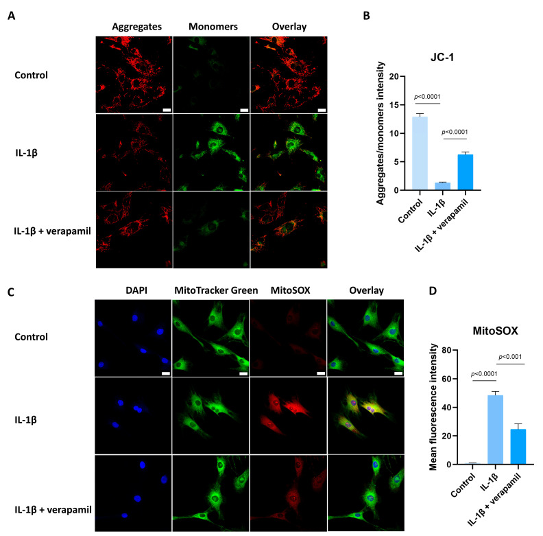
Tendinopathy is a prevalent condition in orthopedics patients, exerting a profound impact on tendon functionality. However, its underlying mechanism remains elusive and the efficacy of pharmacological interventions continues to be suboptimal. Verapamil is a clinically used medicine with anti-inflammation and antioxidant functions. This investigation aimed to elucidate the impact of verapamil in tendinopathy and the underlying mechanisms through which verapamil ameliorates the severity of tendinopathy. In in vitro experiments, primary tenocytes were exposed to interleukin-1 beta (IL-1β) along with verapamil at a concentration of 5 μM. In addition, an in vivo rat tendinopathy model was induced through the localized injection of collagenase into the Achilles tendons of rats, and verapamil was injected into these tendons at a concentration of 5 μM. The in vitro findings highlighted the remarkable ability of verapamil to attenuate extracellular matrix degradation and apoptosis triggered by inflammation in tenocytes stimulated by IL-1β. Furthermore, verapamil was observed to significantly suppress the inflammation-related MAPK/NFκB pathway. Subsequent investigations revealed that verapamil exerts a remediating effect on mitochondrial dysfunction, which was achieved through activation of the Nrf2/HO-1 pathway. Nevertheless, the protective effect of verapamil was nullified with the utilization of the Nrf2 inhibitor ML385. In summary, the in vivo and in vitro results indicate that the administration of verapamil profoundly mitigates the severity of tendinopathy through suppression of inflammation and activation of the Nrf2/HO-1 pathway. These findings suggest that verapamil is a promising therapeutic agent for the treatment of tendinopathy, deserving further and expanded research.

Keywords: Nrf2/HO-1 axis; ROS; mitochondrial dysfunction; tendinopathy; verapamil.


Conflict of interest statement

The authors declare no conflicts of interest.

Figures

Figure 1
Figure 1
Toxicity of verapamil on tenocytes at different concentrations. (A) The effects of verapamil on cell viability were assessed via a CCK8 assay at 24, 48, and 72 h. (B) Live/dead cell staining of tenocytes treated with 0, 0.625, 1.25, 2.5, or 5 μM verapamil. Scale bar, 250 μm. Data are presented in the form of mean ± standard deviation, n = 3. A difference was deemed no significant (ns) if the p-value was greater than 0.05.
Figure 2
Figure 2
Verapamil protects tenocytes from extracellular matrix degradation, inflammation, and apoptosis induced by IL−1β. (A) The relative mRNA expression of IL6, COX2, MMP3, MMP9, and MMP13 analyzed via qPCR. (B) The protein level of BAX, BCL2, IL6, MMP3, MMP9, and MMP13 detected by means of Western blot. (C) Quantitative results of BAX, BCL2, IL6, MMP3, MMP9, and MMP13 detected by means of Western blot. (D) Immunofluorescent images of MMP13 obtained in combination with DAPI staining for the cell nucleus. Scale bar, 100 μm. (E) Quantitative results of MMP13 immunofluorescence. Data are presented in the form of mean ± standard deviation, n = 3.
Figure 3
Figure 3
RNA sequencing and analysis comparing normal tenocytes with those stimulated by IL −1β. (A) Heatmap of genes differentially expressed between normal tenocytes and those stimulated with IL−1β. (B) Volcanol plot of genes differentially expressed between normal tenocytes and those stimulated by IL−1β. (C) Chart of differentially upregulated and downregulated genes between normal tenocytes and those stimulated with IL−1β; genes with |log2FC| > 1 and p-adjust < 0.05 were considered to be significantly different expressed genes. (D) Gene Ontology (GO) enrichment analysis of differentially expressed genes; rich factor is the ratio of differentially expressed protein number annotated in this pathway term to all protein number annotated. (E) Inflammatory activity-, apoptosis-, extracellular matrix degradation-, and oxidative stress-related genes. (F) Kyoto Encyclopedia of Genes and Genomes (KEGG) pathway analysis; rich factor is the ratio of differentially expressed protein numbers annotated in this pathway term to all annotated protein numbers. n = 3.
Figure 4
Figure 4
Expression of the NFκB and MAPK signaling pathways after IL−1β and verapamil administration. (A) The phosphorylation levels of IκBα and P65 in tenocytes of the control, IL−1β, and IL−1β + verapamil groups examined by means of Western blot. (B) Quantitative results of phosphorylation levels of P65 and IκBα and quantitative results of IκBα. (C) The phosphorylation levels of P38 and ERK1/2 in tenocytes of the control, IL−1β, and IL−1β + verapamil groups examined by means of Western blot. (D) Quantitative results of phosphorylation levels of P38 and ERK1/2. Data are presented in the form of mean ± standard deviation, n = 3.
Figure 5
Figure 5
Protective effect of verapamil on IL−1β-induced oxidative stress in tenocytes. (A) DCFH-DA probe and DAPI staining for nucleus. Scale bar, 200 μm. (B) Quantitative results of DCFH-DA. (C) Dihydroethidium probe and DAPI staining for nucleus. Scale bar, 200 μm. (D) Quantitative results of DHE. Data are presented in the form of mean ± standard deviation, n = 3.
Figure 6
Figure 6
Protective effect of verapamil on IL−1β-induced mitochondrial dysfunction in tenocytes. (A) Fluorescent images of JC-1 staining. Scale bar, 20 μm. (B) Quantitative results of JC-1. (C) MitoTracker Green staining for the mitochondria and MitoSOX probe for the superoxide combined with DAPI staining for the nucleus. Scale bar, 20 μm. (D) Quantitative results of MitoSOX. Data are presented in the form of mean ± standard deviation, n = 3.
Figure 7
Figure 7
Verapamil facilitates Nrf2 entry into the nucleus to exert its protective effects. (A) The levels of Nrf2 translocation to the nucleus and the expression level of HO-1 in tenocytes of the control, IL−1β, and IL−1β + verapamil groups examined by Western blot. (B) Quantitative results of Nrf2 and HO-1. (C) DCFH-DA probe and DAPI staining for nucleus among the control, IL−1β, IL−1β + verapamil, and IL−1β + verapamil + 2 μM ML385 groups. Scale bar, 200 μm. (D) Quantitative results of DCFH-DA. Data are presented in the form of mean ± standard deviation, n = 3.
Figure 8
Figure 8
ML385 reverses the protective effect of verapamil. (A) Immunofluorescent images of HO-1 obtained in combination with DAPI staining for the cell nucleus among the control, IL−1β, IL−1β + verapamil, and IL−1β + verapamil + 2 μM ML385 groups. Scale bar, 150 μm. (B) Quantitative results of HO-1 staining among the control, IL−1β, IL−1β + verapamil, and IL−1β + verapamil + 2 μM ML385 groups. (C) Immunofluorescent images of MMP13 obtained in combination with DAPI staining for the cell nucleus among the control, IL−1β, IL−1β + verapamil, and IL−1β + verapamil + 2 μM ML385 groups. Scale bar, 150 μm. (D) Quantitative results of MMP13 staining among the control, IL−1β, IL−1β + verapamil, and IL−1β + verapamil + 2 μM ML385 groups. Data are presented in the form of mean ± standard deviation, n = 3.
Figure 9
Figure 9
ML385 has the capacity to reverse the protective function of verapamil on the mitochondrial membrane potential. (A) Fluorescent images of JC-1 staining among the control, IL−1β, IL−1β + verapamil, and IL−1β + verapamil + 2 μM ML385 groups. (B) Quantitative results of JC-1 staining among the control, IL−1β, IL−1β + verapamil, and IL−1β + verapamil + 2 μM ML385 groups. Scale bar, 20μm. Data are presented in the form of mean ± standard deviation, n = 3.
Figure 10
Figure 10
Evaluation of the protective effect of verapamil on rat tendons in vivo. (A) HE and Masson’s trichrome staining of tendons. Scale bar, 100 μm. (B) Histology score of HE staining among the four groups. Scale bar, 100 μm. (C) TUNEL staining among the four groups. (D) Quantitative results of TUNEL staining. (E) Immunohistochemical staining of MMP13 and COX2 among the four groups. Scale bar, 100 μm. (F) Quantitative results of immunohistochemical staining of MMP13 and COX2. Data are presented in the form of mean ± standard deviation, n = 6.
Figure 11
Figure 11
Evaluation of the protective effect of verapamil on rat tendons in vivo. (A) Immunohistochemical staining of HO-1, P-ERK1/2, P-P38, and P-P65 among the four groups. Scale bar, 100 μm. (B) Quantitative results of immunohistochemical staining of HO-1, P-ERK1/2, P-P38, and P-P65. Data are presented in the form of mean ± standard deviation, n = 6.

Similar articles

Cited by

References

    1. Kaux J.F., Forthomme B., Goff C.L., Crielaard J.M., Croisier J.L. Current opinions on tendinopathy. J. Sports Sci. Med. 2011;10:238–253. - PMC - PubMed
    1. Riel H., Lindstrøm C.F., Rathleff M.S., Jensen M.B., Olesen J.L. Prevalence and incidence rate of lower-extremity tendinopathies in a Danish general practice: A registry-based study. BMC Musculoskelet. Disord. 2019;20:239. doi: 10.1186/s12891-019-2629-6. - DOI - PMC - PubMed
    1. Albers I.S., Zwerver J., Diercks R.L., Dekker J.H., Van den Akker-Scheek I. Incidence and prevalence of lower extremity tendinopathy in a Dutch general practice population: A cross sectional study. BMC Musculoskelet. Disord. 2016;17:16. doi: 10.1186/s12891-016-0885-2. - DOI - PMC - PubMed
    1. Burry H.C. Pathogenesis of some traumatic and degenerative disorders of soft tissue. Aust. N. Z. J. Med. 1978;8((Suppl. S1)):163–167. doi: 10.1111/j.1445-5994.1978.tb04816.x. - DOI - PubMed
    1. Wang J.H., Jia F., Yang G., Yang S., Campbell B.H., Stone D., Woo S.L. Cyclic mechanical stretching of human tendon fibroblasts increases the production of prostaglandin E2 and levels of cyclooxygenase expression: A novel in vitro model study. Connect. Tissue Res. 2003;44:128–133. doi: 10.1080/03008200390223909. - DOI - PubMed

LinkOut - more resources