. 2024 Apr 19;12(4):907.
doi: 10.3390/biomedicines12040907.

Antitumor Efficacy of Arylquin 1 through Dose-Dependent Cytotoxicity, Apoptosis Induction, and Synergy with Radiotherapy in Glioblastoma Models

Affiliations

Antitumor Efficacy of Arylquin 1 through Dose-Dependent Cytotoxicity, Apoptosis Induction, and Synergy with Radiotherapy in Glioblastoma Models

Ann-Shung Lieu et al. Biomedicines. .

Abstract

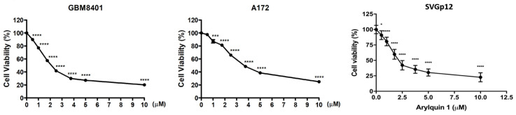
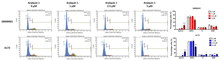
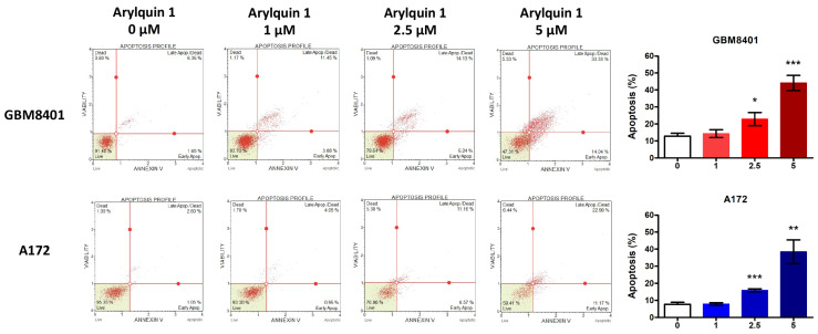
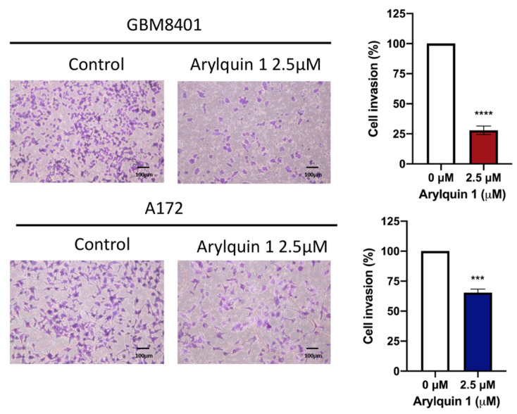
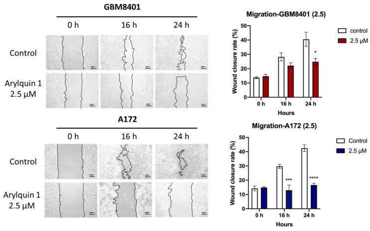
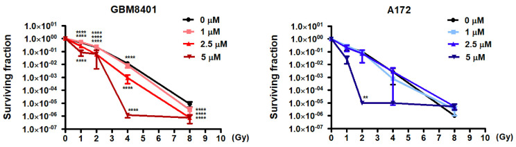
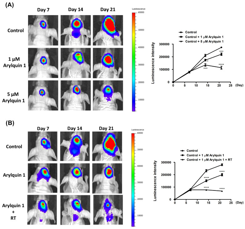
Glioblastoma (GBM), the most aggressive form of brain cancer, is characterized by rapid growth and resistance to conventional therapies. Current treatments offer limited effectiveness, leading to poor survival rates and the need for novel therapeutic strategies. Arylquin 1 has emerged as a potential therapeutic candidate because of its unique mechanism of inducing apoptosis in cancer cells without affecting normal cells. This study investigated the efficacy of Arylquin 1 against GBM using the GBM8401 and A172 cells by assessing its dose-dependent cytotoxicity, apoptosis induction, and synergy with radiotherapy. In vitro assays demonstrated a significant reduction in cell viability and increased apoptosis, particularly at high concentrations of Arylquin 1. Migration and invasion analyses revealed notable inhibition of cellular motility. In vivo experiments on NU/NU nude mice with intracranially implanted GBM cells revealed that Arylquin 1 substantially reduced tumor growth, an effect magnified by concurrent radiotherapy. These findings indicate that by promoting apoptosis and enhancing radiosensitivity, Arylquin 1 is a potent therapeutic option for GBM treatment.

Keywords: Arylquin 1; antitumor efficacy; apoptosis; cytotoxicity; glioblastoma; radiotherapy.


Conflict of interest statement

The authors declare no conflicts of interest.

Figures

Figure 1
Figure 1
Effect of Arylquin 1 on glioblastoma (GBM) cell viability. The graph illustrates the dose-dependent cytotoxicity of Arylquin 1 on GBM8401 and A172 cells. Cell viability was assessed at various concentrations of Arylquin 1 (ranging from 0 to 10 μM). A steep decline in cell viability was observed with increasing concentrations of Arylquin 1, with significant cytotoxic effects noticeable at lower concentrations that became more pronounced with each successive increase in dosage. Statistical significance is indicated by asterisks, with one asterisk (*) denoting p < 0.05, three asterisks (***) denoting p < 0.01, four asterisks (****) denoting p < 0.0001, reflecting a highly significant reduction in cell viability after Arylquin 1 treatment.
Figure 2
Figure 2
Arylquin 1 modulates cell cycle distribution in GBM8401 and A172 cells. Flow cytometry histograms display the DNA content profiles, reflecting the distribution of cells across different phases of the cell cycle in GBM8401 and A172 cells treated with Arylquin 1 at concentrations of 0, 1, 2.5, and 5 μM. Histograms represent the percentage of cells in the G0/G1, S, and G2/M phases, with the Sub G1 peak indicating apoptotic cells. The bar graphs quantify the percentage of cells in each phase of the cell cycle corresponding to each treatment. GBM8401 cells showed an increase in the Sub G1 population with higher doses of Arylquin 1, indicating increased apoptosis. Similarly, A172 cells exhibited a dose-dependent increase in Sub G1, with a marked escalation at 2.5 μM and 5 μM, suggesting enhanced apoptosis. Notable changes in cell cycle phases, such as a decrease in the S and G2/M phases at higher concentrations, indicated cell cycle arrest and reduced proliferation. Asterisks indicate statistical significance compared to the untreated control: * p < 0.05, *** p < 0.001 and **** p < 0.0001.
Figure 3
Figure 3
Arylquin 1 induces dose-dependent apoptosis in GBM8401 and A172 cells. Flow cytometric apoptosis profiles of GBM8401 and A172 cells treated with various concentrations of Arylquin 1 (0, 1, 2.5, and 5 μM). The quadrants represent live (bottom left), early apoptotic (bottom right), late apoptotic/dead (top right), and dead (top left) cells. The corresponding bar graphs summarize the percentage of apoptotic cells, combining the early and late apoptotic populations, for each treatment group. Data indicate a significant increase in apoptosis with higher concentrations of Arylquin 1 in both cells, with GBM8401 showing a notable rise in apoptosis at both at 2.5 μM (* p < 0.05) and 5 μM (*** p < 0.001) and A172 cells displaying a significant increase at both 2.5 μM (** p < 0.01) and 5 μM (*** p < 0.001) compared to the untreated control.
Figure 4
Figure 4
Effects of Arylquin 1 on GBM cell invasiveness in Matrigel invasion assays. Microscopic images depicting the invasive properties of GBM8401 and A172 cells through Matrigel matrices following treatment with Arylquin 1. The left panels show untreated control cells with dense cellular invasion, whereas the right panels show cells post–Arylquin 1 treatment, displaying reduced invasion. The quantified invasion rates for GBM8401 and A172 cells illustrated a significant decline in cell invasion upon Arylquin 1 treatment compared to the controls. Asterisks indicate statistical significance compared to the untreated control: *** p < 0.001 and **** p < 0.0001.
Figure 5
Figure 5
Impact of Arylquin 1 on GBM cell migration in a wound healing essay. Displayed are the results of a wound healing assay performed on GBM8401 and A172 cells to assess the impact of Arylquin 1 on cellular migration. Time-lapse images captured at 0, 8, 12, and 24 h post–Arylquin 1 application documented the changes in wound closure. The images show a noticeable decrease in the wound closure rate of GBM8401 cells after Arylquin 1 treatment compared to that of the control, which was more pronounced at higher concentrations of the drug. A172 cells exhibited a similar reduction in wound-healing capability after treatment. The bar graphs on the right quantitatively depict the percentage of wound closure over time, underlining the inhibitory effect of Arylquin 1 on cell migration in both GBM8401 and A172 cells, with a notable dose-dependent reduction in the migration rate relative to the untreated controls. Asterisks indicate statistical significance compared to the untreated control: * p < 0.05, *** p < 0.001 and **** p < 0.0001.
Figure 6
Figure 6
Enhancing radiosensitivity in GBM cells with Arylquin 1 treatment. The graphs display the surviving fractions of GBM8401 and A172 cells post-irradiation with increasing doses of radiation (0 to 8 Gy) in the presence of varying concentrations of Arylquin 1 (0, 1, 2.5, and 5 μM). For GBM8401 cells, survival decreased with increasing doses of Arylquin 1, indicating a synergistic effect that enhanced radiosensitivity. The surviving fraction was dramatically reduced at the highest concentration of Arylquin 1, particularly at higher radiation doses, as indicated by the steep decline in the red lines. A similar pattern was observed in A172 cells, where Arylquin 1 treatment leads to a substantial decrease in cell survival upon radiation exposure, suggesting increased radiosensitivity. These results demonstrated that Arylquin 1 could potentially be used to sensitize GBM cells to radiation therapy, lowering the surviving fraction of cells more effectively than radiation alone. Statistical significance is denoted by asterisks, indicating a potentiated reduction in survival at each dose level compared to the control. Asterisks indicate statistical significance compared to the untreated control: ** p < 0.01, **** p < 0.0001.
Figure 7
Figure 7
Arylquin 1 impact on tumor growth with and without radiotherapy. (A) The luminescence intensity measurements reveal tumor progression over time in groups treated with different concentrations of Arylquin 1. Compared to the control, both 1 μM and 5 μM doses of Arylquin 1 manifest a clear reduction in tumor growth, with the 5 μM concentration exhibiting a more pronounced suppressive effect. (B) This panel highlights the comparative tumor progression between the control group, a group treated with 1 μM Arylquin 1, and another group receiving a combination of 1 μM Arylquin 1 and radiotherapy (RT). Treatment with 1 μM Arylquin 1 shows a reduction in tumor growth, and, when combined with RT, this effect is significantly amplified, indicating a synergistic interaction that greatly diminishes tumor proliferation. Asterisks indicate statistical significance compared to the untreated control: * p < 0.05, *** p < 0.001 and **** p < 0.0001.

Similar articles

References

    1. Ostrom Q.T., Price M., Neff C., Cioffi G., Waite K.A., Kruchko C., Barnholtz-Sloan J.S. CBTRUS Statistical Report: Primary Brain and Other Central Nervous System Tumors Diagnosed in the United States in 2016–2020. Neuro-Oncology. 2023;25((Suppl. S4)):iv1–iv99. doi: 10.1093/neuonc/noad149. - DOI - PMC - PubMed
    1. Wirsching H.G., Galanis E., Weller M. Glioblastoma. Handb. Clin. Neurol. 2016;134:381–397. - PubMed
    1. Lacroix M., Abi-Said D., Fourney D.R., Gokaslan Z.L., Shi W., DeMonte F., Lang F.F., McCutcheon I.E., Hassenbusch S.J., Holland E., et al. A multivariate analysis of 416 patients with glioblastoma multiforme: Prognosis, extent of resection, and survival. J. Neurosurg. 2001;95:190–198. doi: 10.3171/jns.2001.95.2.0190. - DOI - PubMed
    1. Lamborn K.R., Chang S.M., Prados M.D. Prognostic factors for survival of patients with glioblastoma: Recursive partitioning analysis. Neuro-Oncology. 2004;6:227–235. doi: 10.1215/S1152851703000620. - DOI - PMC - PubMed
    1. Ostrom Q.T., Gittleman H., Farah P., Ondracek A., Chen Y., Wolinsky Y., Stroup N.E., Kruchko C., Barnholtz-Sloan J.S. CBTRUS statistical report: Primary brain and central nervous system tumors diagnosed in the United States in 2006–2010. Neuro-Oncology. 2013;15((Suppl. S2)):ii1–ii56. doi: 10.1093/neuonc/not151. - DOI - PMC - PubMed