. 2024 Apr 17;14(8):1199.
doi: 10.3390/ani14081199.

Effect of High Energy Low Protein Diet on Lipid Metabolism and Inflammation in the Liver and Abdominal Adipose Tissue of Laying Hens

Affiliations

Effect of High Energy Low Protein Diet on Lipid Metabolism and Inflammation in the Liver and Abdominal Adipose Tissue of Laying Hens

Xiaxia Du et al. Animals (Basel). .

Abstract

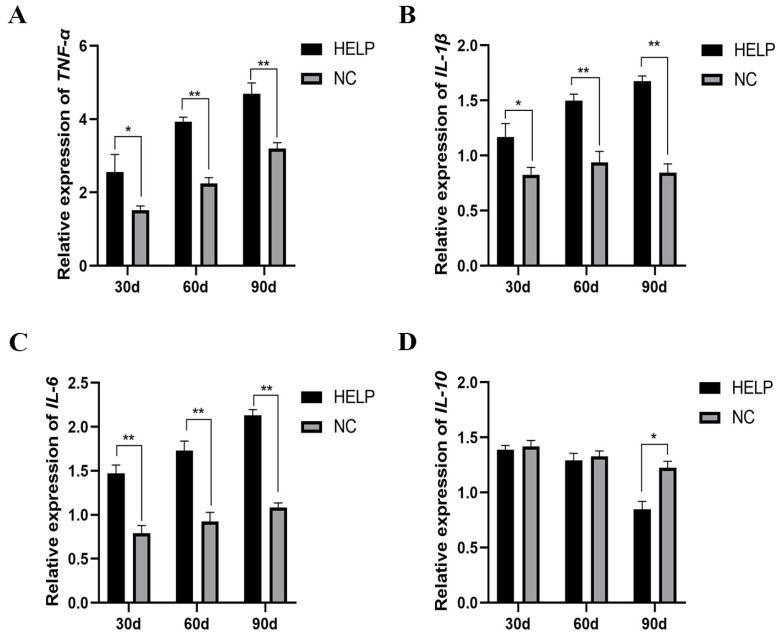
The aim of this study was to evaluate the effects of a high-energy low-protein (HELP) diet on lipid metabolism and inflammation in the liver and abdominal adipose tissue (AAT) of laying hens. A total of 200 Roman laying hens (120 days old) were randomly divided into two experimental groups: negative control group (NC group) and HELP group, with 100 hens per group. The chickens in the NC group were fed with a basic diet, whereas those in the HELP group were given a HELP diet. Blood, liver, and AAT samples were collected from 20 chickens per group at each experimental time point (30, 60, and 90 d). The morphological and histological changes in the liver and AAT were observed, and the level of serum biochemical indicators and the relative expression abundance of key related genes were determined. The results showed that on day 90, the chickens in the HELP group developed hepatic steatosis and inflammation. However, the diameter of the adipocytes of AAT in the HELP group was significantly larger than that of the NC group. Furthermore, the results showed that the extension of the feeding time significantly increased the lipid contents, lipid deposition, inflammatory parameters, and peroxide levels in the HELP group compared with the NC group, whereas the antioxidant parameters decreased significantly. The mRNA expression levels of genes related to lipid synthesis such as fatty acid synthase (FASN), stearoyl-coA desaturase (SCD), fatty acid binding protein 4 (FABP4), and peroxisome proliferator-activated receptor gamma (PPARγ) increased significantly in the liver and AAT of the HELP group, whereas genes related to lipid catabolism decreased significantly in the liver. In addition, the expression of genes related to lipid transport and adipokine synthesis decreased significantly in the AAT, whereas in the HELP group, the expression levels of pro-inflammatory parameters such as tumor necrosis factor-alpha (TNF-α), interleukin-6 (IL-6), and interleukin-1 beta (IL-1β) increased significantly in the liver and AAT. Conversely, the expression level of the anti-inflammatory parameter interleukin-10 (IL-10) decreased significantly in the liver. The results indicated that the HELP diet induced lipid peroxidation and inflammation in the liver and AAT of the laying hens. Hence, these results suggest that chicken AAT may be involved in the development of fatty liver.

Keywords: adipose tissue; fatty liver; inflammation; laying hen; lipid metabolism.


Conflict of interest statement

The authors declare no competing interests.

Figures

Figure 1
Figure 1
Performance and organ weight of the laying hens fed basal and HELP diets for 30, 60, and 90 d. (A) Egg-laying rate. (B) Body weight. (C) Liver weight. (D) AAT weight. Data are expressed as the mean ± SE (n = 20, each group). * p < 0.05, ** p < 0.01.
Figure 2
Figure 2
Morphological changes in the AAT and liver of chickens fed basal and HELP diets for 30, 60, and 90 days. (A) Morphological changes in the chicken liver and AAT. (B) Histological changes in the chicken liver (magnified 400×). (C) Oil Red O staining of frozen slices of the chicken liver (magnified 200×). (D) Histological changes in the adipose tissue of the chicken abdomen (magnified 200×). (E) Comparative analysis of the lipid droplet diameter in the AAT of chickens. Data are expressed as the mean ± SE (n = 6, each group). * p < 0.05, ** p < 0.01.
Figure 3
Figure 3
Transmission electron microscopy of the AAT and liver of the chickens fed with normal and HELP diets for a period of 90 days. (A) Transmission electron microscopy of the chicken liver. (B) Transmission electron microscopy of AAT in chickens. Cell nucleus: n, mitochondria: m, lipid droplets: LD.
Figure 4
Figure 4
Effects of HELP diet on the levels of serum biochemical parameters of laying hens. (A) Total cholesterol, TC. (B) Triglyceride, TG. (C) Alanine aminotransferase, ALT. (D) Aspartate aminotransferase, AST. (E) Total superoxide dismutase, T-SOD. (F) Glutathione, GSH. (G) Tumor necrosis factor-α, TNF-α. (H) Interleukin-6, IL-6. Data are expressed as the mean ± SE (n = 9, each group). * p < 0.05, ** p < 0.01.
Figure 5
Figure 5
Impacts of feeding HELP diet on the liver biochemical parameters of laying hens. (A) Total cholesterol, TC. (B) Triglyceride, TG. (C) Very low-density lipoprotein y, VLDLy. (D) High-density lipoprotein cholesterol, HDL-ch. (E) Malondialdehyde, MDA. (F) Lipid peroxide, LPO. (G) Total superoxide dismutase, T-SOD. (H) Glutathione, GSH. Data are expressed as the mean ± SE (n = 9, each group). * p < 0.05, ** p < 0.01.
Figure 6
Figure 6
The relative expression of genes related to lipolysis, lipid synthesis, and lipid transport in the liver of chickens fed with or without HELP diet. (AC) The mRNA expression of genes related to lipolysis (PPARα, MTTP, and SERBP1). (DF) The mRNA expression of genes related to lipid synthesis (PPARγ, FASN, and SCD). (GI) The expression of genes related to lipid transport (VTGII, ApoB, and LDLR). Data are expressed as the mean ± SE (n = 9, each group). * p < 0.05, ** p < 0.01. Differences marked with ‘ns’ are not statistically significant.
Figure 7
Figure 7
The mRNA expression of genes related to lipid synthesis and hormone synthesis in the AAT of chickens fed with or without HELP diet. The mRNA expression of FABP4 (A), LPL (B), LEPTIN (C), PPARγ (D), CPT-1A (E), and ADPN (F) in the AAT. Data are expressed as the mean ± SE (n = 9, each group). * p < 0.05, ** p < 0.01.
Figure 8
Figure 8
The mRNA expression of genes related to inflammatory parameters in the liver of chickens fed with or without HELP diet. The mRNA expression of TNF-α (A), IL-1β (B), IL-6 (C), IL-10 (D) in the AAT. Data are expressed as the mean ± SE (n = 9, each group). * p < 0.05, ** p < 0.01.
Figure 9
Figure 9
The mRNA expression of genes related to inflammatory parameters in the AAT of chickens fed with or without HELP diet. The mRNA expression of TNF-α (A), IL-1β (B), IL-6 (C) in the AAT. Data are expressed as the mean ± SE (n = 9, each group). * p < 0.05, ** p < 0.01.

References

    1. Harms R.H., Arafa A.S., Simpson C.F., Miles R.D. Influence of Protamone on fatty liver syndrome in commercial laying hens. Poult. Sci. 1982;61:2370–2374. doi: 10.3382/ps.0612370. - DOI - PubMed
    1. O’Hea E.K., Leveille G.A. Lipid biosynthesis and transport in the domestic chick (Gallus domesticus) Comp. Biochem. Physiol. 1969;30:149–159. doi: 10.1016/0010-406X(69)91309-7. - DOI - PubMed
    1. Kartsoli S., Kostara C.E., Tsimihodimos V., Bairaktari E.T., Christodoulou D.K. Lipidomics in non-alcoholic fatty liver disease. World J. Hepatol. 2020;12:436–450. doi: 10.4254/wjh.v12.i8.436. - DOI - PMC - PubMed
    1. Xu Y., Li Z., Zhang S., Zhang H., Teng X. miR-187-5p/apaf-1 axis was involved in oxidative stress-mediated apoptosis caused by ammonia via mitochondrial pathway in chicken livers. Toxicol. Appl. Pharmacol. 2020;388:114869. doi: 10.1016/j.taap.2019.114869. - DOI - PubMed
    1. Simon J., Leroith D. Insulin receptors of chicken liver and brain. Characterization of alpha and beta subunit properties. Eur. J. Biochem. 1986;158:125–132. doi: 10.1111/j.1432-1033.1986.tb09729.x. - DOI - PubMed