

The Impact of Excision Interval on Equine Melanoma Progression: Time Matters?
- PMID: 38672392
- PMCID: PMC11047369
- DOI: 10.3390/ani14081244
The Impact of Excision Interval on Equine Melanoma Progression: Time Matters?
Abstract
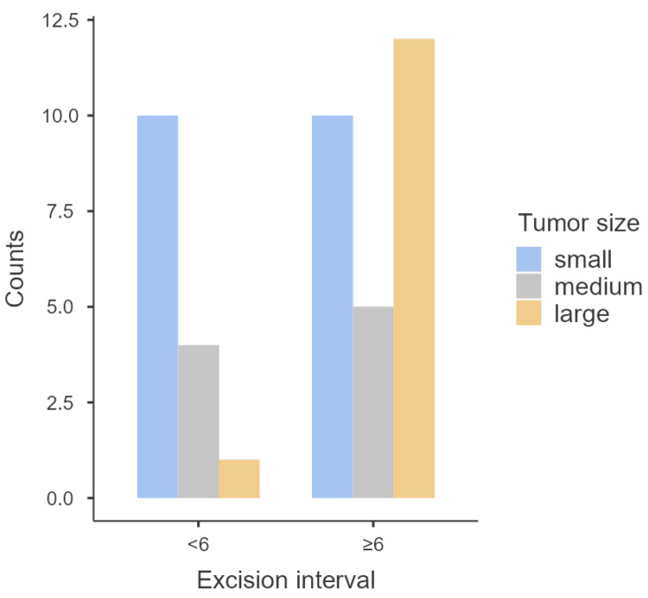
Equine melanomas are a common neoplasm in gray horses. However, scientific knowledge about their progression over time is quite scarce. Some owners and veterinarians still believe that early intervention is not necessary, stating that tumors evolve very slowly and intervention could worsen the animal's condition. This work aims to identify clinical and histological differences that may exist between equine melanomas with different excision intervals (time between tumor detection and surgical excision). A total of 42 tumors (13 benign and 29 malignant) from 34 horses were included in this study. There was a statistically significant association between excision interval and tumor size (p = 0.038), with tumors excised later being significantly larger than the ones excised sooner. The excision interval was also statistically associated with the number of tumors (p = 0.011), since the horses that carried a tumor for longer seemed to be prone to have multiple tumors. Furthermore, there was an association between excision interval and malignancy (p = 0.035), with tumor excised later being fives times more likely to be malignant. This study provides evidence of delayed excision's effect on the progression of equine melanomas. Additionally, it reinforces the importance of the early excision of these tumors.
Keywords: clinical factors; equine; histology; melanomas; time.
Conflict of interest statement
The authors declare no conflicts of interest.
Figures


Similar articles
-
E-Cadherin Immunostaining in Equine Melanocytic Tumors.Animals (Basel). 2023 Jul 6;13(13):2216. doi: 10.3390/ani13132216. Animals (Basel). 2023. PMID: 37444014 Free PMC article.
-
Isolation, establishment, and characterization of ex vivo equine melanoma cell cultures.In Vitro Cell Dev Biol Anim. 2009 Mar-Apr;45(3-4):152-62. doi: 10.1007/s11626-008-9156-3. Epub 2008 Dec 5. In Vitro Cell Dev Biol Anim. 2009. PMID: 19057970
-
Pain severity scores for common equine disorders as provided by horse owners and equine veterinarians.Equine Vet J. 2022 Nov;54(6):1094-1102. doi: 10.1111/evj.13559. Epub 2022 Feb 7. Equine Vet J. 2022. PMID: 35034381
-
Melanomas of the conjunctiva.Int Ophthalmol Clin. 1980 Summer;20(2):161-75. Int Ophthalmol Clin. 1980. PMID: 6995385 Review.
-
"Personalized Excision" of Malignant Melanoma-Need for a Paradigm Shift in the Beginning Era of Personalized Medicine.Am J Dermatopathol. 2019 Dec;41(12):884-896. doi: 10.1097/DAD.0000000000001450. Am J Dermatopathol. 2019. PMID: 31490196 Review.
Cited by
-
Equine Veterinarian Perspectives on Mucocutaneous Tumors in Horses: A Survey-Based Study in Portugal.Animals (Basel). 2025 Jun 23;15(13):1853. doi: 10.3390/ani15131853. Animals (Basel). 2025. PMID: 40646752 Free PMC article.
References
-
- Knottenbelt D.C., Patterson-Kane J.C., Snalune K.L. Clinical Equine Oncology. Elsevier; Amsterdam, The Netherlands: 2015. Melanocytic Neoplasms; pp. 237–246.
-
- Sullins K.E. Melanocytic Tumours in Horses. Equine Vet. Educ. 2020;32:624–630. doi: 10.1111/eve.13159. - DOI
-
- Valentine B.A. The Spectrum of Equine Melanocytic Tumours. Equine Vet. Educ. 2003;15:24. doi: 10.1111/j.2042-3292.2003.tb00206.x. - DOI
Grants and funding
LinkOut - more resources
Full Text Sources
