. 2024 Apr 11;14(4):470.
doi: 10.3390/biom14040470.

Functional Integrity of Radical SAM Enzyme Dph1•Dph2 Requires Non-Canonical Cofactor Motifs with Tandem Cysteines

Affiliations

Functional Integrity of Radical SAM Enzyme Dph1•Dph2 Requires Non-Canonical Cofactor Motifs with Tandem Cysteines

Koray Ütkür et al. Biomolecules. .

Abstract

The Dph1•Dph2 heterodimer from yeast is a radical SAM (RS) enzyme that generates the 3-amino-3-carboxy-propyl (ACP) precursor for diphthamide, a clinically relevant modification on eukaryotic elongation factor 2 (eEF2). ACP formation requires SAM cleavage and atypical Cys-bound Fe-S clusters in each Dph1 and Dph2 subunit. Intriguingly, the first Cys residue in each motif is found next to another ill-defined cysteine that we show is conserved across eukaryotes. As judged from structural modeling, the orientation of these tandem cysteine motifs (TCMs) suggests a candidate Fe-S cluster ligand role. Hence, we generated, by site-directed DPH1 and DPH2 mutagenesis, Dph1•Dph2 variants with cysteines from each TCM replaced individually or in combination by serines. Assays diagnostic for diphthamide formation in vivo reveal that while single substitutions in the TCM of Dph2 cause mild defects, double mutations almost entirely inactivate the RS enzyme. Based on enhanced Dph1 and Dph2 subunit instability in response to cycloheximide chases, the variants with Cys substitutions in their cofactor motifs are particularly prone to protein degradation. In sum, we identify a fourth functionally cooperative Cys residue within the Fe-S motif of Dph2 and show that the Cys-based cofactor binding motifs in Dph1 and Dph2 are critical for the structural integrity of the dimeric RS enzyme in vivo.

Keywords: ADP-ribosylation; Dph1•Dph2; Saccharomyces cerevisiae; cysteine ligands; diphthamide modification; eEF2; iron sulfur cluster; radical SAM enzyme.


Conflict of interest statement

K.M. and U.B. are employed by and members of Roche Pharma Research & Early Development (pRED) and are co-inventors on patent applications that cover assays to detect the presence or absence of diphthamide. Roche is interested in targeted therapies and diagnostics. All other authors declare no conflicts of interest.

Figures

Figure 1
Figure 1
TCM conservation in atypical radical SAM and Fe-S motifs from Dph1•Dph2 dimers. (A) Dph1 and Dph2 alignment from indicated eukaryotic species to archaeal Dph2 (for full details, see Figure S1). (B) Structural comparisons. Left: structure of the PhDph2•Dph2 homodimer (firebrick red; PDB:3LZD) with the Cys-based (Cys-59; Cys-163; Cys-287) Fe-S binding motif previously established [7,9,10,12]. Note Cys-59 is next to Asp-60. Right: an AlphaFold model of the ScDph1•Dph2 heterodimer (forest and lemon green) confirming that cysteines in Dph1 (Cys-133; Cys-239; Cys-368) and Dph2 (Cys-C107; Cys-128; Cys-362) are proximal to respective Fe-S clusters. As part of TCMs, a fourth cysteine, Cys-134 next to Cys-133 in Dph1 and Cys-106 next to Cys-107 in Dph2, orients towards each Fe-S cluster. Fe-S motif close-ups are 66% transparent for emphasis on stick structures. Dph1•Dph2 was modelled and illustrated as previously described [10,21].
Figure 2
Figure 2
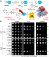
DPH1 and DPH2 mutagenesis to study TCM relevance for Dph1•Dph2 activity in vivo. (A) Simplified diphthamide pathway with DPH1 and DPH2 genes required to initiate ACP formation [21]; subsequent DPH products and steps for the completion of diphthamide synthesis [14,15,16,17] are not detailed. Diphthamide can be hijacked to induce cell death (skull–crossbones) either by DT for lethal ADP-ribosylation of eEF2 or sordarin (sor), which in a complex with the décor on eEF2 stalls ribosomes [40]. (B) Growth assays in response to DT and sordarin for the diagnosis of diphthamide defects in vivo. Yeast tester strains comprised wild-type (WT: DPH1 DPH2) and null mutants (dph1∆; dph2∆) together with single/double DPH1 or DPH2 gene substitutions, as indicated. Cells were grown w/o DT and sordarin (control: left panel), under DT-inducing conditions (+ DT: middle panel) [24], or with sordarin (+ sordarin: right panel) doses sufficient to inhibit WT cells. Arrows shaded in green denote various degrees of DT and sordarin resistance.
Figure 3
Figure 3
Accumulation of unmodified eEF2 in Cys-to-Ser Dph1•Dph2 mutants reveals functional roles of TCMs in diphthamide synthesis. (A) Western blots diagnostic for diphthamide synthesis on eEF2 from cell extracts of indicated yeast strains; anti-eEF2(no diphthamide) antibodies specific for unmodified eEF2 (upper panels) produce immune signals that associate with dysfunctional Dph1•Dph2 rather than WT enzymes, and anti-eEF2(pan) antibodies (middle panels) recognize eEF2 irrespective of modification state [31,32,33]. Technical repetitions (n = 3) were quantified (lower panels) using t-test statistics (* = p < 0.05; ** = p < 0.01; **** = p < 0.0001; n.s. = not significant). (B) ADP-ribosylation (ADPR) assay. Cell extracts incubated w/o (upper panel) or with exotoxin A (ETA) (500 ng, lower panel) with biotin-NAD+ allowed for diphthamide-dependent biotin-ADP-ribose transfer (ADPR-eEF2). Detection was performed via Western blot using a streptavidin-HPR-conjugate. Asterisks mark an unspecific ADPR band previously described [24]. Original images can be found in the Supplementary Materials (Figures S2–S4).
Figure 4
Figure 4
Malfunctional Cys-to-Ser variants of the Dph1•Dph2 dimer have significantly decreased amounts of Dph1 and Dph2 subunits. (A) Western blot detection of Dph1-HA (anti-HA) and Dph2-c-Myc (anti-c-Myc) from total extracts of indicated DPH1-HA substitution mutants co-expressing DPH2-c-Myc (left panel). Detection of Cdc19 (anti-Cdc19) served as internal control. Technical repetitions (n = 3) were followed by densitometric quantification of signal intensities (right panel) and t-test statistics (* = p < 0.05; ** = p < 0.01; *** = p < 0.001; **** = p < 0.0001; n.s. = not significant). (B) Cycloheximide chase of dph1C368S reveals accelerated decay of Dph1•Dph2. Schematic workflow of the cycloheximide chase (left panel). Yeast cells coding for HA and c-Myc-tagged Dph1 and Dph2 were grown to the exponential phase (t0) before the addition of cycloheximide. Samples were taken at indicated time points before (t0) and after cycloheximide addition for protein extraction and Western blot analysis (right panel). Original images can be found in the Supplementary Materials (Figures S5 and S8).

Similar articles

References

    1. Frey P.A., Hegeman A.D., Ruzicka F.J. The radical SAM superfamily. Crit. Rev. Biochem. Mol. Biol. 2008;43:63–88. doi: 10.1080/10409230701829169. - DOI - PubMed
    1. Hoffman B.M., Broderick W.E., Broderick J.B. Mechanism of radical initiation in the radical SAM enzyme superfamily. Annu. Rev. Biochem. 2023;92:333–349. doi: 10.1146/annurev-biochem-052621-090638. - DOI - PMC - PubMed
    1. Yokoyama K., Lilla E.A. C-C bond forming radical SAM enzymes involved in the construction of carbon skeletons of cofactors and natural products. Nat. Prod. Rep. 2018;35:660–694. doi: 10.1039/C8NP00006A. - DOI - PMC - PubMed
    1. Nicolet Y. Structure–function relationships of radical SAM enzymes. Nat. Catal. 2020;3:337–350. doi: 10.1038/s41929-020-0448-7. - DOI
    1. Broderick J.B., Duffus B.R., Duschene K.S., Shepard E.M. Radical S-adenosylmethionine enzymes. Chem. Rev. 2014;114:4229–4317. doi: 10.1021/cr4004709. - DOI - PMC - PubMed

Publication types

MeSH terms