. 2024 Apr 16;16(8):1511.
doi: 10.3390/cancers16081511.

CALR but Not JAK2 Mutations Are Associated with an Overexpression of Retinoid X Receptor Alpha in Essential Thrombocythemia

Affiliations

CALR but Not JAK2 Mutations Are Associated with an Overexpression of Retinoid X Receptor Alpha in Essential Thrombocythemia

Ana Guijarro-Hernández et al. Cancers (Basel). .

Abstract

Essential thrombocythemia (ET) is a blood cancer caused by mutations in JAK2 and CALR. It is widely recognized that both mutations lead to the constitutive activation of JAK2/STAT signaling, although other JAK/STAT-independent pathogenic mechanisms triggered by these alterations have also been described in ET. In an attempt to study JAK2/STAT-independent mechanisms derived from CALR mutations, our research group created a C. elegans model with patient-like mutations in calreticulin that lacks JAK counterparts. The introduction of patient-like mutations in the calreticulin of C. elegans leads to an increase in the transcriptional expression of nhr-2, independently of JAK2/STAT activation. In the present study, we aim to verify if this mechanism is conserved in patients with ET harboring CALR mutations. To do so, we evaluated the expression of potential orthologs of nhr-2 in human cell lines of interest for the study, as well as in bone marrow (BM) or peripheral blood (PB) mononuclear cells from patients with CALR or JAK2 mutations. The results revealed that this mechanism is conserved in CALR-mutated ET patients, since CALR, but not JAK2 mutations, were associated with an overexpression of RXRA in patients with ET. The use of drugs targeting the activation or blockade of this target in the analyzed cell lines did not result in changes in cell viability. However, RXRA might be relevant in the disease, pointing to the need for future research testing retinoids and other drugs targeting RXRα for the treatment of ET patients.

Keywords: CALR; ET; RXRA; myeloproliferative neoplasms.


Conflict of interest statement

A.G.-H., C.H., M.J.L. and J.L.V. declare no conflicts of interest. M.J.C. declares honoraria for lectures from and membership on advisory boards with Janssen, Jazz Pharmaceuticals, Astellas, Novartis, Amgen, and Bristol-Myers Squibb-Celgene.

Figures
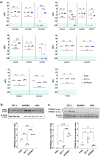
Figure 1
Figure 1
(a) Normalized expression of some of the nhr-2 orthologs in the human cell lines K562, SET-2, and MARIMO. Individual values (n = 3), means, and standard deviations are shown. When no expression was detected in any of the samples analyzed, no statistic is shown (ND, not detected). (b,c) Analysis of RXRα and PPARγ expression by Western Blot in the cell lines K562, SET-2, and MARIMO. The antibody for PPARγ detects both isoforms PPARγ1 and PPARγ2. Individual values (n = 3), means, and standard deviations are shown in the graphs. Differences were considered non-significant (ns) when p > 0.05, significant (*) when p < 0.05, very significant (**) when p < 0.01, and highly significant (***) when p < 0.001.
Figure 2
Figure 2
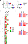
(a) Normalized expression of human nhr-2 orthologs RXRA, RXRB, RXRG, and PPARG in samples from 38 patients with ET and mutations in CALR (26 with type 1 mutations and 12 with type 2 mutations), 21 patients with ET and mutations in JAK2, and 10 healthy donors. Individual values, means, and standard deviations are shown. When no expression was detected in any of the samples analyzed, no statistic is shown (ND, not detected). (b) Individual heatmaps with RXRA, RXRB, RXRG, and PPARG expression data in the samples. Some of the samples were extracted from bone marrow (BM) and others from peripheral blood (PB). Additionally, the time of sample collection is indicated, either at the time of diagnosis or during follow-up of the disease. In all cases, average fold change values are represented from shades of green (negative fold change) to shades of red (positive fold change). (c) The percentage of viable cells quantified by the MTS assay after exposing MARIMO and SET-2 cell lines to increasing concentrations (0, 0.1, 1, and 10 μM) of bexarotene, HX531, and ATRA without ruxolitinib or in combination with ruxolitinib at 1 μM (n = 3). * p < 0.05, ** p < 0.01, and *** p < 0.001.

References

    1. Guijarro-Hernández A., Vizmanos J.L. A broad overview of signaling in Ph-negative classic myeloproliferative neoplasms. Cancers. 2021;13:984. doi: 10.3390/cancers13050984. - DOI - PMC - PubMed
    1. Guijarro-Hernández A., Eder-Azanza L., Hurtado C., Navarro-Herrera D., Ezcurra B., Novo F.J., Cabello J., Vizmanos J.L. Transcriptomic analysis reveals JAK2/MPL-independent effects of calreticulin mutations in a C. elegans model. Cells. 2023;12:186. doi: 10.3390/cells12010186. - DOI - PMC - PubMed
    1. Schneider C.A., Rasband W.S., Eliceiri K.W. NIH Image to ImageJ: 25 years of image analysis. Nat. Methods. 2012;9:671–675. doi: 10.1038/nmeth.2089. - DOI - PMC - PubMed
    1. Quentmeier H., MacLeod R.A., Zaborski M., Drexler H.G. JAK2 V617F tyrosine kinase mutation in cell lines derived from myeloproliferative disorders. Leukemia. 2006;20:471–476. doi: 10.1038/sj.leu.2404081. - DOI - PubMed
    1. Uozumi K., Otsuka M., Ohno N., Moriyama T., Suzuki S., Shimotakahara S., Matsumura I., Hanada S., Arima T. Establishment and characterization of a new human megakaryoblastic cell line (SET-2) that spontaneously matures to megakaryocytes and produces platelet-like particles. Leukemia. 2000;14:142–152. doi: 10.1038/sj.leu.2401608. - DOI - PubMed