Review
. 2024 Apr 19;16(8):1568.
doi: 10.3390/cancers16081568.

Coagulation Protease-Driven Cancer Immune Evasion: Potential Targets for Cancer Immunotherapy

Affiliations
Review

Coagulation Protease-Driven Cancer Immune Evasion: Potential Targets for Cancer Immunotherapy

Subhojit Paul et al. Cancers (Basel). .

Abstract

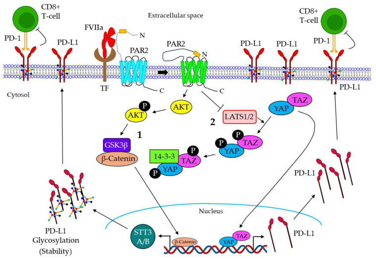
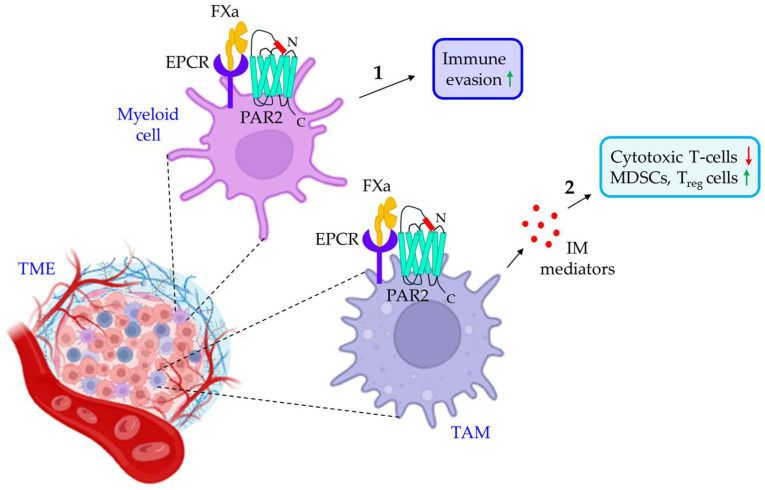
Blood coagulation and cancer are intrinsically connected, hypercoagulation-associated thrombotic complications are commonly observed in certain types of cancer, often leading to decreased survival in cancer patients. Apart from the common role in coagulation, coagulation proteases often trigger intracellular signaling in various cancers via the activation of a G protein-coupled receptor superfamily protease: protease-activated receptors (PARs). Although the role of PARs is well-established in the development and progression of certain types of cancer, their impact on cancer immune response is only just emerging. The present review highlights how coagulation protease-driven PAR signaling plays a key role in modulating innate and adaptive immune responses. This is followed by a detailed discussion on the contribution of coagulation protease-induced signaling in cancer immune evasion, thereby supporting the growth and development of certain tumors. A special section of the review demonstrates the role of coagulation proteases, thrombin, factor VIIa, and factor Xa in cancer immune evasion. Targeting coagulation protease-induced signaling might be a potential therapeutic strategy to boost the immune surveillance mechanism of a host fighting against cancer, thereby augmenting the clinical consequences of targeted immunotherapeutic regimens.

Keywords: biomarker; blood coagulation; cancer; clinical trial; coagulation protease; immune evasion; immunotherapy; protease-activated receptor.


Conflict of interest statement

The authors declare no conflict of interest.

Figures

Figure 1
Figure 1
Mechanism of PAR activation by coagulation proteases. Coagulation proteases cleave PARs at the N-terminal extracellular domain, leading to the generation of a new N-terminus end. This newly formed N-termini acts as a tethered ligand that binds to the ECL2 region of the receptor itself, resulting in the activation of the receptor. Agonist peptides often activate PARs by directly binding to the receptor and do not require PAR cleavage. PAR activation mediated by different coagulation proteases triggers either intracellular G protein- or β-arrestin-induced signaling, leading to different cellular responses. PAR: protease-activated receptor; ECL2: extracellular loop 2.
Figure 2
Figure 2
The role of coagulation protease-driven PAR signaling in cancer. PAR1, PAR2, and PAR4 are shown to be associated with cancer progression. PAR1 signaling in cancer. 1. aPC promotes cancer cell migration via PAR1 activation. Thrombin triggers cancer progression in multiple ways. 2. Thrombin stimulates the PAR1-dependent activation of the C and EGFR pathways, resulting in tumor growth. 3. Thrombin/PAR1 signaling also induces MCP1, MMP-2, and MMP-9 expression, triggering tumor metastasis. 4. Thrombin/PAR1 signaling also promotes tumor angiogenesis via the induction of VEGF and VEGFR. 5–7. The FXa-mediated activation of PAR1 is shown to promote tumor growth and proliferation while down-regulating apoptosis. The role of PAR2 in cancer. 8. TF/FVIIa/PAR2 signaling promotes cancer proliferation via the activation of PKCα and the ERK pathway. 9. The TF/FVIIa-dependent activation of the AKT/GSK3β pathway induces the migration of cancer cells. 10. TF/FVIIa/PAR2 signaling also induces tumor metastasis via the AKT/NF-ĸB pathway. 11. The TF/FVIIa-mediated activation of PAR2 promotes the release of miR-221-laden MVs from metastatic breast cancer (BC) cells, which deliver miR-221 to non-metastatic BC cells, thereby inducing proliferation, metastasis, and anti-apoptosis to MV-fused recipient cells. 12. FXa also stimulates PAR2 to promote tumor growth. PAR4 signaling in cancer. 13. Thrombin stimulates PAR4 to induce cancer cell proliferation via the activation of the ERK, p38, and AKT signaling pathways. The green upward arrows indicate up-regulation; the red downward arrows indicate down-regulation. PAR: protease-activated receptor: aPC: activated protein C: NF-ĸB: nuclear factor kappa-light-chain-enhancer of activated B cells: EGFR: extracellular growth factor receptor, MCP1: monocyte chemoattractant protein-1; MMP: matrix metalloproteinase; VEGF: vascular endothelial growth factor; VEGFR: VEGF receptor; FXa: activated factor X; TF: tissue factor; FVIIa: activated factor VII; PKCα: protein kinase Cα; ERK: extracellular signal-regulated kinase; GSK3β: glycogen synthase kinase 3β; miR: microRNA.
Figure 3
Figure 3
Different types of cancer immune evasion mechanisms. 1. Cancer cells are either devoid of antigens or remove antigens, thereby down-regulating tumor immunogenicity. 2. Tumor cells inhibit DC maturation via the release of MCSF, IL-10, prostaglandin, VEGF, TGF-β, and IDO. 3. In the TME, cells exhibit lower expression of co-stimulatory molecules, leading to decreased T-cell activity. 4. Cancer cells inhibit chemokines and secrete VEGF and IL-10, which together down-regulate the infiltration of T-cells. 5. Tumor cells exhibit a reduced expression of MHC-I, proteasome components, β2-microglobulin, and TAP1/2, thereby avoiding immune recognition. 6. In the TME, macrophages are converted to M2-macrophages, which promote the release of IL-10, triggering the down-regulation of the CD8+ T-cell response. 7. In the TME, MDSCs release TGF-β, which down-regulates CD8+ T-cell activity. MDSC also induces Treg cells. Together, these perturb the CD8+ T-cell response. 8. Tumor cells release IDO, which is converted to kynurenine; this further reduces the activity of cytotoxic T-cells, induces MDSC and Treg cells, and, ultimately, leads to the down-regulation of the CD8+ T-cell response. The Red down arrows indicate down-regulation; the green up arrows indicate up-regulation. MCSF: macrophage colony-stimulating factor; IL: interleukin; VEGF: vascular endothelial growth factor; TGF-β: transforming growth factor β; IDO: indoleamine 2:3 dioxygenase; DC: dendritic cell; VEGF: vascular endothelial growth factor; IL: interleukin; MHC-I: major histocompatibility complex I; TAP: transporter associated with antigen processing; MDSC: myeloid-derived suppressor cells; Treg cells: regulatory T-cells.
Figure 4
Figure 4
The role of thrombin in cancer immune evasion. 1. In the TME, thrombin triggers the cleavage of GARP on platelets, leading to the release of TGF-β1 from the GARP-LTGF-β1 complex. The released TGF-β1 binds to the receptor on cytotoxic T-cells, thereby conferring immunosuppressive functions. 2. Thrombin also suppresses anti-tumor immunity. 3. Furthermore, thrombin is shown to promote the evasion of cytotoxic T-cells. 4. Thrombin stimulates the release of IL-6 from various cells in the TME, which down-regulates the differentiation, maturation, and antigen-presenting ability of DC. 5. In the TME, thrombin is also shown to induce the release of TNF-α, which not only increases the accumulation and activity of Treg cells, Breg cells, and MDSCs but also evades the cytotoxic T-cell response via down-regulating infiltration and up-regulating apoptosis. 6. The thrombin-mediated release of MCP-1 in the TME also triggers macrophage differentiation into M2 phenotypes, leading to immunosuppressive responses. The green upward arrows indicate induction; the red downward arrows indicate inhibition. GARP: glycoprotein A repetitions predominant; LTGF-β1: latent TGF-β1; IL: interleukin; DC: dendritic cell; Ag: antigen; TNF-α: tumor necrosis factor α; MDSC: myeloid-derived suppressor cells; Treg cells: regulatory T-cells; Breg cells: regulatory B-cells; MCP-1: monocyte chemoattractant protein 1.
Figure 5
Figure 5
Mechanism of FVIIa-mediated cancer immune evasion. 1. The TF/FVIIa-mediated activation of PAR2 promotes AKT phosphorylation, which triggers the GSK3β-dependent accumulation of β-catenin into the nucleus, resulting in the induction of STT3A/B expression in TNBC cells. The induced expression of STT3A/B promotes PD-L1 glycosylation, leading to the stability of PD-L1. 2. TF/FVIIa/PAR2 signaling also promotes the inactivation of LATS1/2, which is essential for YAP/TAP phosphorylation and their retention in the cytosol. LATS1/2 inactivation results in the nuclear translocation of YAP/TAZ, leading to the induction of PD-L1. PD-L1 is translocated on the surface of the cancer cells, which further binds the PD-1 receptor of CD8+ T-cells, leading to T-cell inhibition. FVIIa: active factor VII; TF: tissue factor; GSK3β: glycogen synthase kinase 3 beta; PD-L1: programmed death-ligand 1; PD-1: programmed cell death protein 1; CD: cluster of differentiation; LATS1/2: large tumor suppressor kinase ½; YAP: yes-associated protein; TAZ: Tafazzin.
Figure 6
Figure 6
The role of FXa/PAR2 signaling in cancer immune evasion. 1. FXa in the TME myeloid cells triggers PAR2 to promote tumor immune evasion. 2. TAMs in the TME also express PAR2, which is activated by FXa, leading to the release of IM mediators. IM mediators promote tumor immune evasion by either reducing the population of cytotoxic T-cells or up-regulating MDSCs and Treg cells. The green upward arrows indicate up-regulation; the red downwards arrows indicate down-regulation. TME: tumor microenvironment; EPCR: endothelial cell protein C receptor; FXa: activated factor X; PAR2: protease-activated receptor 2; TAM: tumor-associated macrophage; IM: immunomodulatory; MDSCs: myeloid-derived suppressor cells; Treg cells: regulatory T-cells.

Similar articles

Cited by

References

    1. Hemker H.C., Kahn M.J. Reaction sequence of blood coagulation. Nature. 1967;215:1201–1202. doi: 10.1038/2151201a0. - DOI - PubMed
    1. Dahlback B. Blood coagulation. Lancet. 2000;355:1627–1632. doi: 10.1016/S0140-6736(00)02225-X. - DOI - PubMed
    1. Walsh P.N., Ahmad S.S. Proteases in blood clotting. Essays Biochem. 2002;38:95–111. doi: 10.1042/bse0380095. - DOI - PubMed
    1. Rezaie A.R. Protease-activated receptor signalling by coagulation proteases in endothelial cells. Thromb. Haemost. 2014;112:876–882. doi: 10.1160/th14-02-0167. - DOI - PMC - PubMed
    1. Heuberger D.M., Schuepbach R.A. Protease-activated receptors (PARs): Mechanisms of action and potential therapeutic modulators in PAR-driven inflammatory diseases. Thromb. J. 2019;17:4. doi: 10.1186/s12959-019-0194-8. - DOI - PMC - PubMed

LinkOut - more resources