. 2024 Apr 11;14(4):494.
doi: 10.3390/life14040494.

Evodiamine Alleviates 2,4-Dinitro-1-Chloro-Benzene-Induced Atopic Dermatitis-like Symptoms in BALB/c Mice

Affiliations

Evodiamine Alleviates 2,4-Dinitro-1-Chloro-Benzene-Induced Atopic Dermatitis-like Symptoms in BALB/c Mice

So-Young Han et al. Life (Basel). .

Abstract

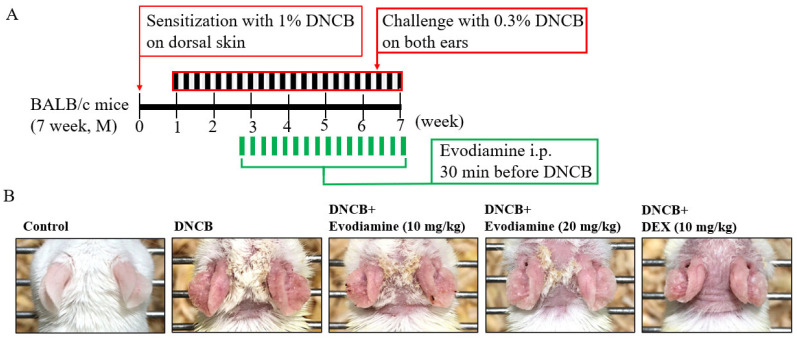
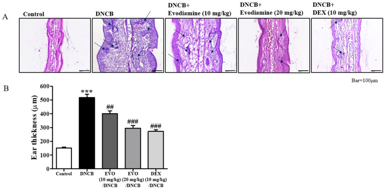
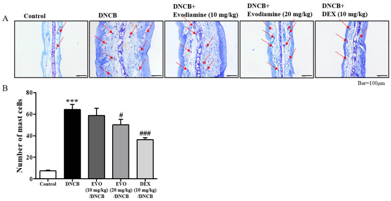
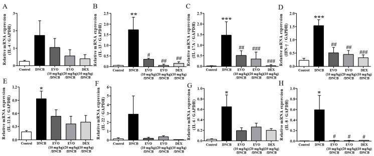
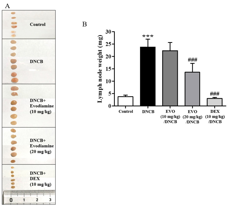
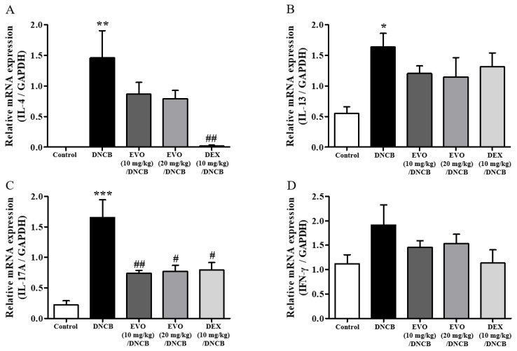
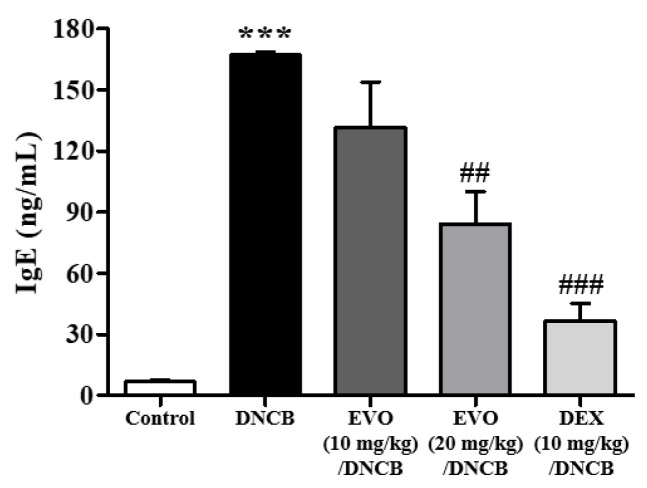
Evodiamine is an alkaloid found in Evodia fruits, a traditional Chinese medicine. Preclinical studies have demonstrated its anti-inflammatory and neuroprotective properties. The 2,4-dinitro-1-chloro-benzene (DNCB) was used to test the effects of evodiamine on a chemically induced atopic dermatitis-like model in BALB/c mice. Evodiamine significantly lowered serum immunoglobulin E levels, which increased as an immune response to the long-term application of DNCB. Several atopic dermatitis-like skin symptoms induced by DNCB, including skin thickening and mast cell accumulation, were suppressed by evodiamine therapy. DNCB induced higher levels of pro-inflammatory cytokines in type 2 helper T (Th2) cells (IL-4 and IL-13), Th1 cells (IFN-γ and IL-12A), Th17 cells (IL-17A), Th22 cells (IL-22), and chemokines (IL-6 and IL-8). These increases were suppressed in the lymph nodes and skin following evodiamine treatment. The results of our study indicate that evodiamine suppresses atopic dermatitis-like responses in mice and may therefore be useful in treating these conditions.

Keywords: Evodia rutaecarpa; anti-atopy; atopy; dermatitis; eczema; evodiamine.


Conflict of interest statement

The authors declare no conflicts of interest.

Figures

Figure 1
Figure 1
Effect of evodiamine on DNCB-induced atopic dermatitis in the ears. Evaluation of the effect of evodiamine on atopic dermatitis induced by DNCB in ears. (A) An outline of the protocol for induction of atopic dermatitis and treatment with evodiamine. On day 0, DNCB was applied to the skin to sensitize it. Following repeated DNCB challenges on days 7–48, atopic dermatitis-like phenotypes were induced. In addition to DNCB, vehicles were applied topically to BALB/c mice, while evodiamine and DEX were injected intraperitoneally 30 min before DNCB exposure (n = 5). (B) A view of the ear in its entirety.
Figure 2
Figure 2
Effect of evodiamine on ear thickness. These are representative histological findings from cutaneous tissue sections taken on day 49. In order to stain the ear tissue, hematoxylin and eosin stain was applied (A). Blue arrows indicated immune cells. A comparison of the ear thickness between the groups was made in (B). Five mice were used for each group. Statistical significance: *** p < 0.001 vs. the vehicle-treated control group; ### p < 0.001, ## p < 0.01 vs. the DNCB-treated group. A magnification of 200× was used.
Figure 3
Figure 3
Effect of evodiamine on mast cell count in the ears. In order to identify mast cells, the skin was stained with toluidine blue O. (A) Histological findings on day 49 of representative cutaneous tissue sections. Mast cells are indicated with red arrows. (B) The number of mast cells in the ear tissues is shown in the histogram (n = 5). Statistical significance: *** p < 0.001 vs. the vehicle-treated control group; ### p < 0.001, # p < 0.05 vs. the DNCB-treated group. A magnification of 200× was used.
Figure 4
Figure 4
Effect of evodiamine on pro-inflammatory cytokine expression in the ears. Based on mouse ear tissue mRNA isolated from the ears of mice, qRT-PCR analysis of the Th2 cytokines IL-4 (A) and IL-13 (B) cytokines, Th17 cytokine, IL-17A (C), and Th1 cytokine, INF-γ (D) and IL-12A (E), Th22 cytokine, IL-22 (F), and chemokines IL-6 (G) and IL-8 (H) was conducted (n = 5). Statistical significance: *** p < 0.001, ** p < 0.01, * p < 0.05 vs. the vehicle-treated control group; ### p < 0.001, ## p < 0.01, # p < 0.05 vs. the DNCB-treated group. Normalization was also performed by comparing mRNA levels to GAPDH mRNA levels.
Figure 5
Figure 5
Effect of evodiamine on the size of lymph nodes. (A) Photographs were taken of the lymph nodes to measure their morphological changes. (B) A measurement of lymph node weight was also conducted (n = 5). Statistical significance: *** p < 0.001 vs. the vehicle-treated control group; ### p < 0.001 vs. the DNCB-treated group.
Figure 6
Figure 6
Effect of evodiamine on the expression of pro-inflammatory cytokines in lymph nodes. Based on mouse lymph node tissue mRNA isolated from the lymph nodes of mice, qRT-PCR analysis of the Th2 cytokines IL-4 (A) and IL-13 (B) cytokines, Th17 cytokine, IL-17A (C), and Th1 cytokine, IFN-γ (D) was conducted (n = 5). A normalization was also performed by comparing mRNA levels to GAPDH mRNA levels. Statistical significance: *** p < 0.001, ** p < 0.01, * p < 0.05 vs. the vehicle-treated control group; ## p < 0.01, # p < 0.05 vs. the DNCB-treated group.
Figure 7
Figure 7
Effect of evodiamine on serum immunoglobulin E levels. Day 49 was the day on which serum was collected from the animals. An enzyme-linked immunosorbent assay was used to measure serum IgE levels (n = 5). Statistical significance: *** p < 0.001 vs. the vehicle-treated control group; ### p < 0.001, ## p < 0.01 vs. the DNCB-treated group.

Similar articles

Cited by

References

    1. Labib A., Yosipovitch G. An evaluation of abrocitinib for moderate-to-severe atopic dermatitis. Expert Rev. Clin. Immunol. 2022;18:1107–1118. doi: 10.1080/1744666X.2022.2126353. - DOI - PubMed
    1. Sroka-Tomaszewska J., Trzeciak M. Molecular Mechanisms of Atopic Dermatitis Pathogenesis. Int. J. Mol. Sci. 2021;22:4130. doi: 10.3390/ijms22084130. - DOI - PMC - PubMed
    1. Yang G., Seok J.K., Kang H.C., Cho Y.Y., Lee H.S., Lee J.Y. Skin Barrier Abnormalities and Immune Dysfunction in Atopic Dermatitis. Int. J. Mol. Sci. 2020;21:2867. doi: 10.3390/ijms21082867. - DOI - PMC - PubMed
    1. Silverberg J.I., Barbarot S., Gadkari A., Simpson E.L., Weidinger S., Mina-Osorio P., Rossi A.B., Brignoli L., Saba G., Guillemin I. Atopic dermatitis in the pediatric population: A cross-sectional, international epidemiologic study. Ann. Allergy Asthma Immunol. 2021;126:417–428.e412. doi: 10.1016/j.anai.2020.12.020. - DOI - PubMed
    1. Wollenberg A., Barbarot S., Bieber T., Christen-Zaech S., Deleuran M., Fink-Wagner A., Gieler U., Girolomoni G., Lau S., Muraro A., et al. Consensus-based European guidelines for treatment of atopic eczema (atopic dermatitis) in adults and children: Part I. J. Eur. Acad. Dermatol. Venereol. 2018;32:657–682. doi: 10.1111/jdv.14891. - DOI - PubMed

LinkOut - more resources