Review
. 2024 Mar 29;14(4):368.
doi: 10.3390/jpm14040368.

Association of Fetal Lung Development Disorders with Adult Diseases: A Comprehensive Review

Affiliations
Review

Association of Fetal Lung Development Disorders with Adult Diseases: A Comprehensive Review

Alexey V Yaremenko et al. J Pers Med. .

Abstract

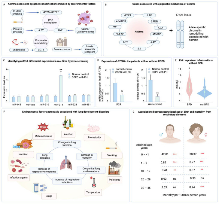
Fetal lung development is a crucial and complex process that lays the groundwork for postnatal respiratory health. However, disruptions in this delicate developmental journey can lead to fetal lung development disorders, impacting neonatal outcomes and potentially influencing health outcomes well into adulthood. Recent research has shed light on the intriguing association between fetal lung development disorders and the development of adult diseases. Understanding these links can provide valuable insights into the developmental origins of health and disease, paving the way for targeted preventive measures and clinical interventions. This review article aims to comprehensively explore the association of fetal lung development disorders with adult diseases. We delve into the stages of fetal lung development, examining key factors influencing fetal lung maturation. Subsequently, we investigate specific fetal lung development disorders, such as respiratory distress syndrome (RDS), bronchopulmonary dysplasia (BPD), congenital diaphragmatic hernia (CDH), and other abnormalities. Furthermore, we explore the potential mechanisms underlying these associations, considering the role of epigenetic modifications, transgenerational effects, and intrauterine environmental factors. Additionally, we examine the epidemiological evidence and clinical findings linking fetal lung development disorders to adult respiratory diseases, including asthma, chronic obstructive pulmonary disease (COPD), and other respiratory ailments. This review provides valuable insights for healthcare professionals and researchers, guiding future investigations and shaping strategies for preventive interventions and long-term care.

Keywords: adult respiratory diseases; epigenetic modifications; fetal respiratory diseases; lung development; neonatal outcomes; preventive interventions; transgenerational effects.


Conflict of interest statement

The authors declare no conflicts of interest.

Figures

Figure 4
Figure 4
Overview of fetal lung diseases, their associations with adult respiratory conditions, and long-term health impacts on different infant groups. (A) Association of fetal lung diseases with adult respiratory diseases and lung disorders [18,121]. RDS—respiratory distress syndrome, BPD—bronchopulmonary dysplasia, COPD—chronic obstructive pulmonary disease, CDH—congenital diaphragmatic hernia, CLM—congenital lung malformations. (B) Number of infections in very preterm (VeryPT), late preterm (LatePT), early term (EarlyT), and late term (LateT) infants per month during their first year after birth (the study was carried out in Austria). Adopted from [123] © 2019 PLOS ONE. (C) Correlations between BPD in infancy and following long-term health disorders (the study was carried out in South Korea). PDA—patent ductus arteriosus, LRI—lower respiratory illness [126]. (D) Comparative analysis of reactive airway disease in infants with BPD, matched cohort controls (infants who were cared for in the intensive care nursery but did not receive mechanical ventilation, did not have BPD and major congenital anomalies, and lived nearby), and normal controls (healthy infants). The study shows that a large portion of individuals with BPD in infancy, specifically 52 percent, had reactive airway disease. This was significantly higher compared to the normal control group (the study was carried out in the USA) [107]. (E) Results of pulmonary function tests in subjects with BPD in infancy, matched cohort controls, and normal controls (the study was carried out in the USA). PEFR—peak expiratory flow rate, FVC—forced vital capacity. FEY1—forced expiratory volume in one second, FEF25-75—forced expiratory flow between 25 and 75 percent of vital capacity, Vmax50—maximal expiratory flow at 50 percent of vital capacity, FRC—functional residual capacity, TLC—total lung capacity, RV/TLC—the ratio of residual volume to total lung capacity, DLCO—diffusing capacity for carbon monoxide [107]. (F) Association between CDH in infancy and its long-term impact on children’s health (the study was carried out in Japan) [127].
Figure 1
Figure 1
Fetal lung development: stages, influential factors, and its pivotal role in postnatal health. (A) Stages and key points of human lung development: The different stages and important aspects of human lung development are shown in diagrams. These stages include embryonic, pseudoglandular, canalicular, saccular, and alveolar. During the embryonic stage, primary branches and SOX2 (Transcription Factor SOX-2)/SOX9 (Transcription Factor SOX-9) co-expression in the tips are visible. Ongoing tip SOX2/SOX9 co-expression and airway differentiation are depicted in the pseudoglandular stage. The canalicular stage shows SOX9+/SOX2 distal tips, and alveolar differentiation begins. In the saccular stage, distal tips disappear and alveolar differentiation progresses, with the expression of SOX9 (cartilage), SOX2 (airway cells), and ACTA2 (Actin Alpha 2, Smooth Muscle). The postnatal alveolar stage shows continued growth and septal formation, with the expression of NKX2-1 (NK2 Homeobox 1) (lung epithelium), FOXF1 (Forkhead Box F1) (mesenchyme), and ACTA2. The SOX9+ distal tips are no longer visible at this stage. Cryosections are reproduced from [19]. Text descriptions are adapted from [33]. (B) Early-life exposures can affect respiratory disease development. Maternal factors like diet, smoking, and medication use can impact fetal immune programming. Early-life lung colonization is crucial for immune response shaping. Changes in lung development and exposure can lead to respiratory diseases later in life. Abnormal lung structure from preterm birth or chorioamnionitis-associated bronchopulmonary dysplasia can increase susceptibility to respiratory complications [30]. IL: interleukin; ST: suppression of tumorigenicity; ILC: innate lymphoid cell; Th: T-helper; PH: pulmonary hypertension. (C) Influence of oxygen level alterations on lung development: Both low and high oxygen levels can lead to vascular dysmorphogenesis, which increases the risk of wheezing and asthma in children [34].
Figure 2
Figure 2
Schematic illustration and brief description of main fetal lung development disorders. (A) Respiratory distress syndrome (RDS), which is characterized by surfactant deficiency and labored breathing [91,92]. (B) Bronchopulmonary dysplasia (BPD), which is linked to premature birth and persistent lung injury [13,17]. (C) Congenital diaphragmatic hernia (CDH), where abdominal organs migrate into the thoracic cavity impeding pulmonary growth) [23,26], and (D) other fetal lung abnormalities including congenital pulmonary airway malformation (CPAM), Pulmonary Sequestration, Congenital Lobar Emphysema (CLE), and pulmonary hypoplasia (PH) [97,98,99,100].
Figure 3
Figure 3
Timeline of human lung development and main factors affecting this process. The development of the lungs is a continuous process with overlapping stages, as most processes begin at the center and progress towards the periphery. The timing of microvascular maturation and the completion of alveolarization is unclear, resulting in fading bars. The embryonic period is not exclusively dedicated to lung development [21]. The risk factors for lung diseases are visually presented in a graph, categorized by the different stages of life: in utero and perinatal life, early childhood, and adulthood [120]. COPD—chronic obstructive pulmonary disease, BPD—bronchopulmonary dysplasia, RDS—respiratory distress syndrome.
Figure 5
Figure 5
Interplay of epigenetic mechanisms in lung development and disease susceptibility. (A) Basic epigenetic mechanisms associated with lung development, illustrating cellular processes including DNA methylation, histone modification, and microRNA (miRNA) regulation within a cell. DNMT—DNA methyltransferase, HAT—histone acetyltransferase, HDAC—histone deacetyltransferase, HDT—histone demethyltransferase, HMT—histone methyltransferase, MV—microvesicle, TET—10–11 translocation methylcytosine dioxygenase [162]. (B) Epigenetic susceptibility to pulmonary diseases, highlighting the impact of gender, nutrition, environmental toxins, pollutants, stress, and parental smoking from birth through adulthood, alongside associated methylation patterns and transcription activities for normal, COPD, and asthma conditions. HOX5—hypermethylation of homeobox 5, LMO2—LIM domain only 2, IL-10—interleukin 10, SMAD3—mothers against decapentaplegic homologue 3 [162]. (C) Pathogenesis of idiopathic pulmonary fibrosis, delineating the roles of various factors in healthy and injured alveoli, the influence of imbalanced histone deacetylase activities on lung fibrosis progression, and the cellular and molecular responses involved in this process. ECM—extracellular matrix, HDAC—histone deacetylase, FMD—fibroblast-to-myofibroblast differentiation, ROS—reactive oxygen species, AECII—type-I/-II alveolar epithelial cell, SASP—senescence-associated secretory phenotype, HAT—histone acetyltransferase, Me—methylation, P—phosphorylation, Ac—acetylation [164,165].
Figure 6
Figure 6
Epigenetic and environmental determinants of lung development. (A) Epigenetic modifications from environmental factors like smoking and ozone on gene expression related to asthma. GSTM1—glutathione S-transferase Mu 1, GSTP1—glutathione S-transferase P1, IL1R—interleukin-1 receptor, TNF—tumor necrosis factor [168]. (B) Key genes implicated in asthma through epigenetic changes [168]. (C) miRNA profiles in COPD patients with pulmonary hypertension (PH), suggesting their regulatory impact and diagnostic potential. Adopted from [169]. (D) Altered expression of the PTEN gene in COPD-associated PH, indicating epigenetic modulation. Adopted from [169]. (E) Comparison of lung changes in preterm infants with and without bronchopulmonary dysplasia (BPD). EML—epigenetic mutation load. Adopted from [170]. (F) Spectrum of environmental influences on lung development disorders [171,172,173,174,175,176,177,178,179,180,181,182]. (G) Correlation between gestational age at birth and respiratory disease mortality rates, highlighting epigenetic sensitivity during early development [183]. Asterisks indicate * p < 0.05, ** p < 0.01, *** p < 0.001, ns—non-significant: p > 0.05.
Figure 7
Figure 7
Strategies and outcomes in fetal lung development interventions. (A) Therapeutic strategies for managing fetal lung development disorders, including mechanical ventilation and CPAP, alongside the administration of surfactants and corticosteroids to enhance lung maturity and function [40,41]. (B) The impact of antenatal corticosteroid exposure on neonatal outcomes such as RDS, showing adjusted odds and beneficial effects of the intervention [198]. AC—antenatal corticosteroids, NAC—no antenatal corticosteroids, RSD—respiratory distress syndrome, vs.—ventilatory support, TT—transient tachypnea, DR—resuscitation in the delivery room, Composite—respiratory distress syndrome, transient tachypnea of the newborn, ventilatory support. (C) Various respiratory support outcomes, including the use of CPAP/INSURE and mechanical ventilation, on respiratory distress in newborns [199]. CPAP/INSURE—a bubble CPAP system, Oxygen/MV—oxygen via nasal cannula, RSD—respiratory distress syndrome, MV—mechanical ventilation, SR—surfactant requirement, Pth—pneumothorax, BPD—bronchopulmonary dysplasia. (D) The effectiveness of CPAP in managing respiratory syndrome across different age groups, demonstrating a significant reduction in respiratory rates compared to control groups. Adopted from [200] © 2017 Elsevier Ltd. (E) Covariates associated with pulmonary function in adolescents, highlighting the influence of environmental and lifestyle factors, as well as allergic diseases on lung function at age 15. Adopted from [201]. (F) Lung function trajectories from childhood to adult life, depicting potential catch-up in lung function and factors influencing normal and low growth trajectories, emphasizing the importance of early-life interventions in respiratory health. Adopted from [202] © 2017 F1000Research. Asterisks indicate * p < 0.05, ** p < 0.01, *** p < 0.001.
Figure 8
Figure 8
Stages of lung development influenced by Wnt signaling. Embryonic stage—HOX5 in the mesenchyme directs Wnt2/2b in the endoderm to form lung progenitors and initiate smooth muscle and cartilage development. Pseudoglandular stage—lung branching morphogenesis is driven by mesenchymal Wnt5a and regulated by Notum, with Wnt7b-BMP4 axis aiding in tissue growth. Canalicular/Saccular stages—Wnt signaling, modulated by DKK1, differentiates the lung epithelium, while high Wnt levels promote distal airway formation. Alveolar stage—Wnt-responsive ATII cells guide the maturation of alveolar structures. Adapted from [229].

References

    1. He P., Lim K., Sun D., Pett J.P., Jeng Q., Polanski K., Dong Z., Bolt L., Richardson L., Mamanova L., et al. A Human Fetal Lung Cell Atlas Uncovers Proximal-Distal Gradients of Differentiation and Key Regulators of Epithelial Fates. Cell. 2022;185:4841–4860.e25. doi: 10.1016/j.cell.2022.11.005. - DOI - PubMed
    1. Cao S., Feng H., Yi H., Pan M., Lin L., Zhang Y.S., Feng Z., Liang W., Cai B., Li Q., et al. Single-Cell RNA Sequencing Reveals the Developmental Program Underlying Proximal–Distal Patterning of the Human Lung at the Embryonic Stage. Cell Res. 2023;33:421–433. doi: 10.1038/s41422-023-00802-6. - DOI - PMC - PubMed
    1. Danopoulos S., Shiosaki J., Al Alam D. FGF Signaling in Lung Development and Disease: Human Versus Mouse. Front. Genet. 2019;10:444859. doi: 10.3389/fgene.2019.00170. - DOI - PMC - PubMed
    1. Loering S., Cameron G.J.M., Starkey M.R., Hansbro P.M. Lung Development and Emerging Roles for Type 2 Immunity. J. Pathol. 2019;247:686–696. doi: 10.1002/path.5211. - DOI - PubMed
    1. Andreas A., Maloy A., Nyunoya T., Zhang Y., Chandra D. The FoxP1 Gene Regulates Lung Function, Production of Matrix Metalloproteinases and Inflammatory Mediators, and Viability of Lung Epithelia. Respir. Res. 2022;23:281. doi: 10.1186/s12931-022-02213-4. - DOI - PMC - PubMed

LinkOut - more resources