. 2024 Apr 10;13(8):2175.
doi: 10.3390/jcm13082175.

Expression of Immunoglobulin G4 in Eosinophilic Esophagitis

Affiliations

Expression of Immunoglobulin G4 in Eosinophilic Esophagitis

See-Young Lee et al. J Clin Med. .

Abstract

Background: Eosinophilic esophagitis (EoE) is a disease that has been subcategorized into two endoscopic phenotypes: inflammatory and fibrostenotic. Moreover, studies have shown a link between EoE and immunoglobulin G4 (IgG4), a subclass of the immunoglobulin G (IgG) antibody. In this study, we aimed to evaluate the relationship between histologic IgG4 expression and endoscopic phenotypes in patients with EoE. Methods: This case-control study included patients diagnosed with EoE (n = 19) and patients with non-obstructive dysphagia without abnormal findings as controls (NOD; n = 12). The EoE group was further divided into three subgroups based on endoscopic phenotype: inflammatory, fibrostenotic, or combined. Retrospective examination of endoscopic findings and pathological slides was performed to analyze IgG4 staining. Results: Histological analysis revealed a significant difference in IgG4 cell count (15.00 vs. 0.58, p = 0.003) and eosinophil cell count (84.67 vs. 0.08, p < 0.001) between the EoE and NOD groups. Symptom manifestation and blood test results were similar across all three endoscopic EoE phenotypes. However, histological analysis revealed a significant difference in IgG4 cell count between the inflammatory, fibrostenotic, and combined phenotypes (4.13 vs. 17.6 vs. 59.7, p = 0.030). Conclusions: IgG4 expression was higher in EoE patients than in those with NOD, the highest being in the combined phenotype subgroup. These findings emphasize the important role of endoscopic and histological examination in diagnosing EoE and the need for further research in this area.

Keywords: EoE endoscopic phenotype; eosinophilic esophagitis; immunoglobulin G.


Conflict of interest statement

The authors declare no conflicts of interest.

Figures

Figure 1
Figure 1
Flowchart of the study participants. Patients complaining of dysphagia were classified into the EoE or NOD groups based on biopsy results. Patients with EoE were divided into inflammatory, combined, or fibrostenotic phenotypes based on their endoscopy results. In this process, one patient with EoE was excluded from phenotype classification owing to normal endoscopy findings. EoE, eosinophilic esophagitis; NOD, non-obstructive dysphagia.
Figure 2
Figure 2
Representative histological analysis of IgG4 and IgG. (A) Example of IgG4 immunostaining shows IgG4 plasma cells (arrow) in the subepithelial area. (B) The ratio of IgG4 to IgG is calculated by counting the number of IgG plasma cells at the same location (9/228 × 100 = 3.94%). Ig, immunoglobulin.
Figure 3
Figure 3
Representative histological analysis of esophageal tissue. (A) Hematoxylin and eosin-stained slide. (B) TRC-stained slide. TRC staining is used to visualize collagen-driven fibrosis, which appears blue in the image. The presence of blue bundles in the TRC-stained slide indicates advanced fibrosis (note that storiform fibrosis, which is characteristic of IgG4-related disease, is not observed in this patient). TRC, Masson’s trichrome.
Figure 4
Figure 4
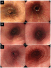
Representative endoscopic analysis of EoE. (A) Inflammatory phenotype. Left image from pre-treatment endoscopy reflects an EREFS of 3 (exudate score of 1 and furrow score of 2). Right image from post-treatment endoscopy reflects an EREFS of 1 (exudate score of 0 and furrow score of 1). The ΔEREFS for this patient is 2. (B) Fibrostenotic phenotype. Left image from pre-treatment endoscopy reflects an EREFS of 2 (ring score of 2). Right image from post-treatment endoscopy reflects an EREFS of 1 (ring score of 1). The ΔEREFS for this patient is 1. (C) Combined phenotype. Left image from pre-treatment endoscopy reflects an EREFS of 3 (furrow score of 1 and ring score of 2). Right image from post-treatment endoscopy reflects an EREFS of 3 (furrow score of 1 and a ring score of 2). The ΔEREFS for this patient is 0. EoE, eosinophilic esophagitis; EREFS, Endoscopic Reference Score; ΔEREFS, change in EREFS.
Figure 5
Figure 5
Analysis results based on EoE phenotype. (A) The graph provides a comparative analysis of serum IgG4 concentrations (mg/L) and IgG4 cell count per HPF across three EoE phenotypes: inflammatory, fibrostenotic, and combined. Using the analysis of variance (ANOVA) method for statistical evaluation, a discernible trend was observed in serum IgG4 levels across the phenotypes (p = 0.077). Moreover, a statistically significant difference was identified in IgG4 cell counts/HPF between the phenotypes (p = 0.030). (B) This graph provides IgG4 cell counts/HPF, TRC positivity rates, and pre-treatment EREFS across EoE phenotypes analyzed using ANOVA. Significant variability in IgG4 cell counts/HPF was noted across phenotypes (p = 0.030), with the combined phenotype showing significantly higher counts. TRC positivity rates also showed significant differences by phenotype (p = 0.028), with both the fibrostenotic and combined phenotypes achieving 100% positivity. Furthermore, variations in pre-treatment EREFS (p = 0.034) were observed, with the combined phenotype displaying higher scores, which may suggest more severe disease manifestations.

References

    1. Furuta G.T., Liacouras C.A., Collins M.H., Gupta S.K., Justinich C., Putnam P.E., Bonis P., Hassall E., Straumann A., Rothenberg M.E. Eosinophilic esophagitis in children and adults: A systematic review and consensus recommendations for diagnosis and treatment. Gastroenterology. 2007;133:1342–1363. doi: 10.1053/j.gastro.2007.08.017. - DOI - PubMed
    1. Park H. Eosinophilic esophagitis. Korean J. Gastroenterol. 2007;50:286–291. - PubMed
    1. Gómez-Aldana A., Jaramillo-Santos M., Delgado A., Jaramillo C., Lúquez-Mindiola A. Eosinophilic esophagitis: Current concepts in diagnosis and treatment. World J. Gastroenterol. 2019;25:4598–4613. doi: 10.3748/wjg.v25.i32.4598. - DOI - PMC - PubMed
    1. Furuta G.T., Katzka D.A. Eosinophilic esophagitis. N. Engl. J. Med. 2015;373:1640–1648. doi: 10.1056/NEJMra1502863. - DOI - PMC - PubMed
    1. Kanakala V., Lamb C.A., Haigh C., Stirling R.W., Attwood S.E. The diagnosis of primary eosinophilic oesophagitis in adults: Missed or misinterpreted? Eur. J. Gastroenterol. Hepatol. 2010;22:848–855. doi: 10.1097/MEG.0b013e32832c7709. - DOI - PubMed

LinkOut - more resources