. 2024 Apr 11;25(8):4218.
doi: 10.3390/ijms25084218.

Entamoeba histolytica: In Silico and In Vitro Oligomerization of EhHSTF5 Enhances Its Binding to the HSE of the EhPgp5 Gene Promoter

Affiliations

Entamoeba histolytica: In Silico and In Vitro Oligomerization of EhHSTF5 Enhances Its Binding to the HSE of the EhPgp5 Gene Promoter

Salvador Pérez-Mora et al. Int J Mol Sci. .

Abstract

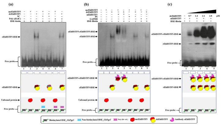
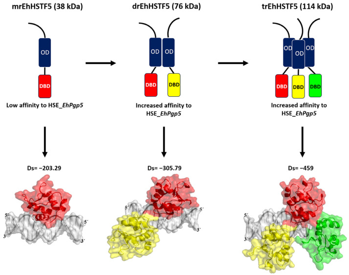
Throughout its lifecycle, Entamoeba histolytica encounters a variety of stressful conditions. This parasite possesses Heat Shock Response Elements (HSEs) which are crucial for regulating the expression of various genes, aiding in its adaptation and survival. These HSEs are regulated by Heat Shock Transcription Factors (EhHSTFs). Our research has identified seven such factors in the parasite, designated as EhHSTF1 through to EhHSTF7. Significantly, under heat shock conditions and in the presence of the antiamoebic compound emetine, EhHSTF5, EhHSTF6, and EhHSTF7 show overexpression, highlighting their essential role in gene response to these stressors. Currently, only EhHSTF7 has been confirmed to recognize the HSE as a promoter of the EhPgp5 gene (HSE_EhPgp5), leaving the binding potential of the other EhHSTFs to HSEs yet to be explored. Consequently, our study aimed to examine, both in vitro and in silico, the oligomerization, and binding capabilities of the recombinant EhHSTF5 protein (rEhHSTF5) to HSE_EhPgp5. The in vitro results indicate that the oligomerization of rEhHSTF5 is concentration-dependent, with its dimeric conformation showing a higher affinity for HSE_EhPgp5 than its monomeric state. In silico analysis suggests that the alpha 3 α-helix (α3-helix) of the DNA-binding domain (DBD5) of EhHSTF5 is crucial in binding to the major groove of HSE, primarily through hydrogen bonding and salt-bridge interactions. In summary, our results highlight the importance of oligomerization in enhancing the affinity of rEhHSTF5 for HSE_EhPgp5 and demonstrate its ability to specifically recognize structural motifs within HSE_EhPgp5. These insights significantly contribute to our understanding of one of the potential molecular mechanisms employed by this parasite to efficiently respond to various stressors, thereby enabling successful adaptation and survival within its host environment.

Keywords: EhDBD5; Entamoeba histolytica; HSE_EhPgp5; dimer; molecular docking; monomer; oligomerization; rEhHSTF5; trimer.


Conflict of interest statement

The authors declare no conflicts of interest.

Figures

Figure 1
Figure 1
Induction and immunodetection of the oligomeric states of the rEhHSTF5 protein. The integrity of the total proteins extracted from transformed and IPTG-induced bacteria was evaluated by SDS-PAGE. A Molecular Weight Marker (MWM) and IPTG induction times ranging from 0 to 24 h were included. The gel was stained with Coomassie Brilliant Blue G-250 to visualize the protein bands (a). Subsequently, immunodetection was performed by Western blot assays using α6xHis and αEhHSTF5 specific antibodies to detect the distinct conformations of rEhHSTF5 (b). The graphs display the relative protein expression, calculated as the average of the pixel values obtained from three independent samples. Expression was normalized using the positive control, αGAPDH. The statistical analysis was performed using a two-way ANOVA, followed by the Tukey’s multiple comparison test. Significant differences between experimental groups are denoted with (*). The significance levels used were ** p ≤ 0.002, and *** p ≤ 0.001.
Figure 2
Figure 2
Purification and immunodetection of oligomeric conformations of the rEhHSTF5 protein. The oligomeric conformations of the rEhHSTF5 protein were monitored before and after loading onto the HisTrap FF column. The total proteins obtained from the bacteria, transformed with the plasmid construct and induced with IPTG (24 h), were passed through filters with a pore size of 0.22 μm (flowthrough, FT), and the proteins that did not bind to the column (waste, W) are shown (a). The chromatogram and SDS-PAGE assays revealed that the drEhHSTF5 protein was eluted in fractions 7 to 12, while the mrEhHSTF5 protein was eluted in fractions 24 to 26 (bd). These proteins were detected using α6xHis and αEhHSTF5 antibodies in WB assays (e,f). In the chromatogram (b), the four increasing imidazole steps are illustrated: the first corresponds to a concentration of 20 mM, followed by the second with 50–100 mM, the third between 200–300 mM, and the fourth at 500 mM, while the blue line indicates the absorbance reading at 280 nm during the elution time, signifying the presence of proteins eluted from the affinity column. For Panels (cf), “F” denotes the fraction number obtained from the chromatography, and “MWM” indicates the molecular weight marker.
Figure 3
Figure 3
mrEhHSTF5 oligomerization induction with increasing concentrations of glutaraldehyde. We used the mrEhHSTF5 protein without glutaraldehyde as a negative control. Oligomerization states were evaluated by 12% SDS-PAGE gel electrophoresis stained with Coomassie Brilliant Blue G-250. The statistical analysis was performed using a two-way ANOVA, followed by the Tukey’s multiple comparison test. Significant differences between experimental groups are denoted with (*). The significance levels used were * p ≤ 0.033, ** p ≤ 0.002, and *** p ≤ 0.001.
Figure 4
Figure 4
HSE recognition of the EhPgp5 gene by the rEhHSTF5 protein. EMSA assays were performed using the monomeric and dimeric conformations of the rEhHSTF5 protein. A specific competitor (non-biotinylated HSE_EhPgp5) and a nonspecific competitor (Poly(dI-dC)) were used (a). For the supershift assays, the specific antibody αEhHSTF5 was utilized for both conformations of the rEhHSTF5 protein. Unrelated antibodies, including mouse preimmune serum (PS) and the αGAPDH antibody, were also used as negative controls (b). Likewise, a kinetic study with increasing drEhHSTF5 concentration (from 0.7 to 2.8 µM) drEhHSTF5 was performed in supershift assays (c). As a negative control, only the biotinylated probe without protein was used. In the image legend, the plus sign (+) indicates the presence of a component in the reaction, while the minus sign (−) denotes its absence. A graphical illustration of each condition used in the EMSA and supershift tests is provided at the bottom.
Figure 5
Figure 5
Construction of 3D models. The 3D structures of HSE_EhPgp5 (a), mEhDBD5 (b), and dEhDBD5 (c) were obtained. Additionally, the representation DBD in the factor EhHSTF5 is illustrated, covering everything from the amino acid sequence to its secondary and three-dimensional structures. The representation includes both the monomeric conformation and the potential homodimeric conformation of the domain. The primary and secondary structures of EhDBD5 are displayed using a color code to facilitate understanding, while the corresponding graph illustrates its stability, assessed in silico through the normalized B-factor provided by the I-TASSER server.
Figure 6
Figure 6
Validation of 3D structures. The 3D models mEhDBD5 (a) and dEhDBD5 (b) were validated using Ramachandran analysis, which provides detailed information about the distribution of dihedral angles of amino acids in favorable and unfavorable regions. The capital letters A, B, and L are used to designate the Most Favored Regions, while the lowercase letters a, b, l, and p represent the Additional Allowed Regions. The tilde (~a, ~b, ~l, ~p) is utilized to indicate the Generously Allowed Regions. Furthermore, a value of −1 is assigned to a torsion angle located within a non-permissible region. The models were subjected to the ProSA-web server for a validation based on the Z-score (the black dot on the graph represents the Z-score assigned to our 3D structure) and global energy, where values ≤ 0 indicate absence of structural errors. ERRAT was used for error quantification, where an asterisk (*) indicates the error value. White bars in the graph represent instances without structural errors, while yellow bars indicate amino acids with structural errors falling within the 95–99% error range, and red bars identify errors exceeding 99%. Verifying 3D structures validates protein structures by evaluating their 3D conformation through comparison to a set of experimental structures. It assigns a score, suggesting an acceptable average 3D−1D score of ≥0.1 for validation.
Figure 7
Figure 7
Molecular docking and intermolecular evaluation of the mEhDBD5-HSE_EhPgp5 and dEhDBD5-HSE_EhPgp5 complexes. Blind dockings were performed with the domains mEhDBD5 or dEhDBD5 and the HSE_EhPgp5. Using the PLIP server, intermolecular interactions of amino acids involved in the interaction with the sense and anti-sense strands of HSE were estimated. A close-up of the area of interaction is shown in the boxes, where amino acids from mEhDBD5 (EhDBD5(1)) are illustrated in red, while the analogue of the second EhDBD5 domain from dEhDBD5 (EhDBD5(2)) is shown in yellow (a,b). The amino acids involved in the interaction with the amino acids of the mEhDBD7-HSE, KlHSTF-HSE, HsHSTF1-HSE, and HsHSTF2-HSE complexes were compared (c). The high conservation degree of α2-helix and α3-helix of DBDs from 28 species, including the EhHSTFs family, is shown (d). In the alignment with the Clustal Omega server, asterisks (*) indicate amino acids that are identical across all sequences. Colons (:) represent conserved substitutions with similar properties, and a period (.) points out semi-conserved substitutions with some functional similarity. The analysis of the physicochemical properties of the complexes revealed that the dEhDBD5-HSE_EhPgp5, containing an additional monomer compared to the mEhDBD5-HSE_EhPgp5, demonstrated more significant aromatic interactions, hydrogen bonds, and charges. This complex also exhibited an increased hydrophobicity and a higher ionization capacity. Furthermore, it possessed a more extensive solvent-exposed surface area than its monomeric counterpart, mEhDBD5-HSE_EhPgp5 (Supplementary Figure S3).
Figure 8
Figure 8
The 3D structural similarity between DBDs–HSEs complexes. Based on the obtained dEhDBD5-HSE_EhPgp5 complex (a), overlay comparisons were performed with crystallographic structures deposited in the PDB database. These included the K. lactis DBD-HSE complex (ID: 3HTS) (b), S. cerevisiae-C. thermophilum complex (ID: 5D5X) (c), H. sapiens-C. thermophilum complex (ID: 5D5W) (d), synthetic H. sapiens construction (ID: 5D8L) (e), and H. sapiens (ID: 7DCU) (f). A joint overlay of all these structures was performed (g) and focusing solely on the wing domain (h). In addition, a significant degree of conservation was identified through the alignment of amino acid sequences of helices 2 and 3 (i). The squares illustrate the TM-scores in percentages, while the RMSD is in Angstroms (Å). In the alignment with the Clustal Omega server, asterisks (*) indicate amino acids that are identical across all sequences. Colons (:) represent conserved substitutions with similar properties, and a period (.) points out semi-conserved substitutions with some functional similarity.
Figure 9
Figure 9
Our in vitro findings demonstrate that the mrEhHSTF5 protein is capable of oligomerizing, forming dimers and trimers. Furthermore, through EMSA assays and in silico molecular docking, we discovered that the dimeric conformation exhibits greater affinity for HSE_EhPgp5 compared to the monomeric conformation. Dorantes et al. [28] reported higher affinity (−459) binding of tEhHSTF5 to the same HSE, supporting our results and indicating that the degree of oligomerization increases the affinity for HSE_EhPgp5. OD; Oligomerization Domain, DBD; DNA Binding Domain, Ds; Docking Score.

Similar articles

Cited by

References

    1. Salles J.M., Moraes L.A., Salles M.C. Hepatic Amebiasis. Braz. J. Infect. Dis. 2003;7:96–110. doi: 10.1590/S1413-86702003000200002. - DOI - PubMed
    1. Varghese V., Kansal A., Bhardwaj S., Sharma A. Cerebral Amebiasis: An Uncommon Cerebral Abscess. Ann. Indian Acad. Neurol. 2021;24:445–447. doi: 10.4103/aian.AIAN_583_20. - DOI - PMC - PubMed
    1. Chou A., Austin R.L. StatPearls. StatPearls Publishing; Treasure Island, FL, USA: 2023. Entamoeba Histolytica Infection. - PubMed
    1. Pineda E., Perdomo D. Entamoeba Histolytica under Oxidative Stress: What Countermeasure Mechanisms Are in Place? Cells. 2017;6:44. doi: 10.3390/cells6040044. - DOI - PMC - PubMed
    1. Uribe-Querol E., Rosales C. Immune Response to the Enteric Parasite Entamoeba Histolytica. Physiology. 2020;35:244–260. doi: 10.1152/physiol.00038.2019. - DOI - PubMed

MeSH terms

LinkOut - more resources