

Altered Glycolysis, Mitochondrial Biogenesis, Autophagy and Apoptosis in Peritoneal Endometriosis in Adolescents
- PMID: 38673823
- PMCID: PMC11050237
- DOI: 10.3390/ijms25084238
Altered Glycolysis, Mitochondrial Biogenesis, Autophagy and Apoptosis in Peritoneal Endometriosis in Adolescents
Abstract
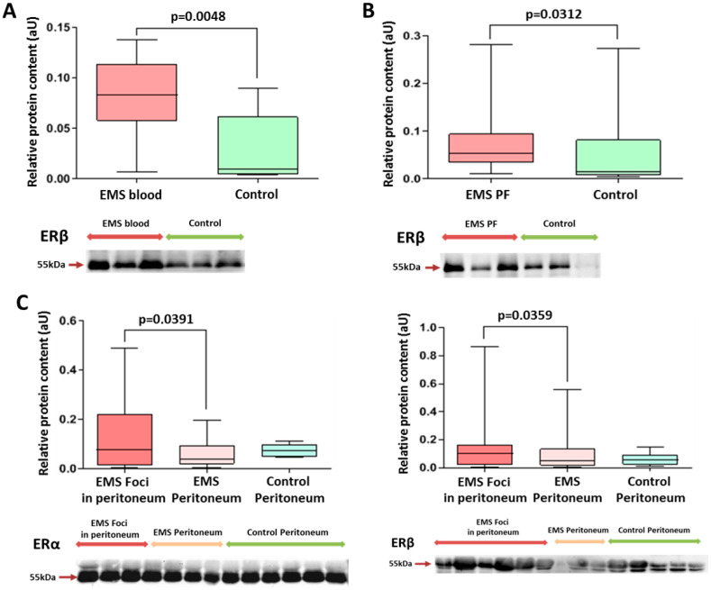
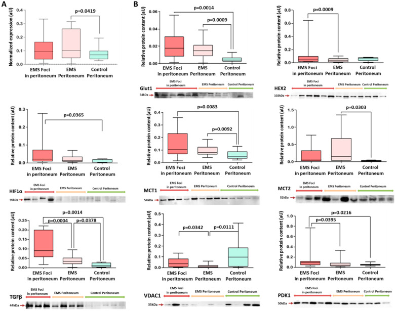
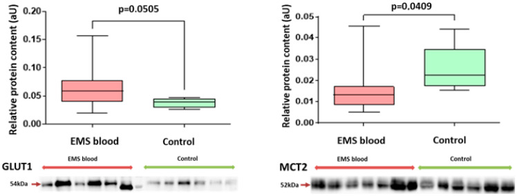
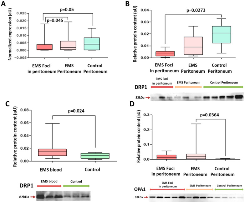
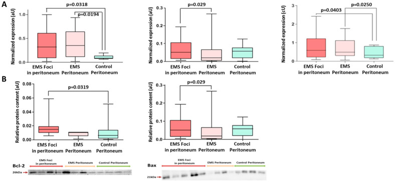
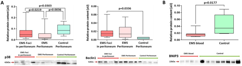
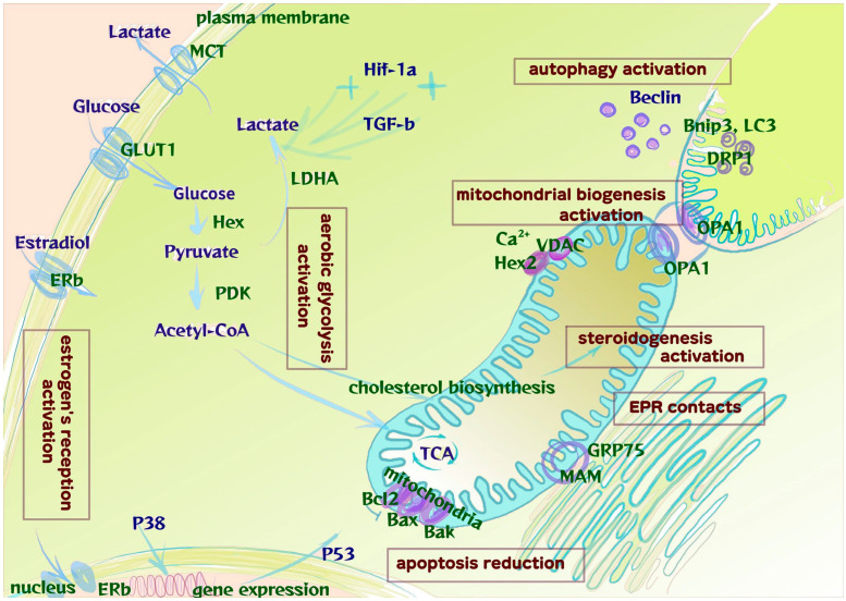
Energy metabolism plays a pivotal role in the pathogenesis of endometriosis. For the initial stages of the disease in adolescents, this aspect remains unexplored. The objective of this paper was to analyze the association of cellular and endosomal profiles of markers of glycolysis, mitochondrial biogenesis, apoptosis, autophagy and estrogen signaling in peritoneal endometriosis (PE) in adolescents. We included 60 girls aged 13-17 years in a case-control study: 45 with laparoscopically confirmed PE (main group) and 15 with paramesonephric cysts (comparison group). Samples of plasma and peritoneal fluid exosomes, endometrioid foci and non-affected peritoneum were tested for estrogen receptor (Erα/β), hexokinase (Hex2), pyruvate dehydrogenase kinase (PDK1), glucose transporter (Glut1), monocarboxylate transporters (MCT1 and MCT2), optic atrophy 1 (OPA1, mitochondrial fusion protein), dynamin-related protein 1 (DRP1, mitochondrial fission protein), Bax, Bcl2, Beclin1, Bnip3, P38 mitogen-activated protein kinase (MAPK), hypoxia-inducible factor 1 (Hif-1α), mitochondrial voltage-dependent anion channel (VDAC) and transforming growth factor (TGFβ) proteins as markers of estrogen signaling, glycolysis rates, mitochondrial biogenesis and damage, apoptosis and autophagy (Western-Blot and PCR). The analysis identified higher levels of molecules associated with proliferation (ERβ), glycolysis (MCT2, PDK1, Glut1, Hex2, TGFβ and Hif-1α), mitochondrial biogenesis (OPA1, DRP1) and autophagy (P38, Beclin1 and Bnip3) and decreased levels of apoptosis markers (Bcl2/Bax) in endometrioid foci compared to non-affected peritoneum and that in the comparison group (p < 0.05). Patients with PE had altered profiles of ERβ in plasma and peritoneal fluid exosomes and higher levels of Glut1, MCT2 and Bnip3 in plasma exosomes (p < 0.05). The results of the differential expression profiles indicate microenvironment modification, mitochondrial biogenesis, estrogen reception activation and glycolytic switch along with apoptosis suppression in peritoneal endometrioid foci already in adolescents.
Keywords: Bcl-2; Hif-1α; TGFβ; adolescents; apoptosis; autophagy; estrogen receptor β; exosomes; glycolysis; mitochondrial biogenesis; peritoneal endometriosis; proliferation.
Conflict of interest statement
The authors declare no conflicts of interest. The funders had no role in the design of the study; in the collection, analyses, or interpretation of data; in the writing of the manuscript; or in the decision to publish the results.
Figures
Similar articles
-
Mitochondrial translocation of estrogen receptor β affords resistance to oxidative insult-induced apoptosis and contributes to the pathogenesis of endometriosis.Free Radic Biol Med. 2019 Apr;134:359-373. doi: 10.1016/j.freeradbiomed.2019.01.022. Epub 2019 Jan 24. Free Radic Biol Med. 2019. PMID: 30684560
-
Transforming growth factor-β induced Warburg-like metabolic reprogramming may underpin the development of peritoneal endometriosis.J Clin Endocrinol Metab. 2014 Sep;99(9):3450-9. doi: 10.1210/jc.2014-1026. Epub 2014 May 5. J Clin Endocrinol Metab. 2014. PMID: 24796928 Free PMC article.
-
17β-Estradiol and/or Estrogen Receptor β Attenuate the Autophagic and Apoptotic Effects Induced by Prolonged Hypoxia Through HIF-1α-Mediated BNIP3 and IGFBP-3 Signaling Blockage.Cell Physiol Biochem. 2015;36(1):274-84. doi: 10.1159/000374070. Epub 2015 May 4. Cell Physiol Biochem. 2015. PMID: 25967966
-
Organelle-specific autophagy in inflammatory diseases: a potential therapeutic target underlying the quality control of multiple organelles.Autophagy. 2021 Feb;17(2):385-401. doi: 10.1080/15548627.2020.1725377. Epub 2020 Feb 12. Autophagy. 2021. PMID: 32048886 Free PMC article. Review.
-
Impaired autophagy and APP processing in Alzheimer's disease: The potential role of Beclin 1 interactome.Prog Neurobiol. 2013 Jul-Aug;106-107:33-54. doi: 10.1016/j.pneurobio.2013.06.002. Epub 2013 Jul 1. Prog Neurobiol. 2013. PMID: 23827971 Review.
Cited by
-
Warburg-like Metabolic Reprogramming in Endometriosis: From Molecular Mechanisms to Therapeutic Approaches.Pharmaceuticals (Basel). 2025 May 28;18(6):813. doi: 10.3390/ph18060813. Pharmaceuticals (Basel). 2025. PMID: 40573210 Free PMC article. Review.
-
Endometriosis in Adolescence: A Narrative Review of the Psychological and Clinical Implications.Diagnostics (Basel). 2025 Feb 24;15(5):548. doi: 10.3390/diagnostics15050548. Diagnostics (Basel). 2025. PMID: 40075795 Free PMC article. Review.
References
-
- Yeung P., Gupta S., Gieg S. Endometriosis in Adolescents: A Systematic Review. J. Endometr. Pelvic Pain Disord. 2017;9:17–29. doi: 10.5301/je.5000264. - DOI
-
- Khashchenko E.P., Uvarova E.V., Chuprynin V.D., Pustynnikova M.Y., Fatkhudinov T.K., Elchaninov A.V., Gardanova Z.R., Ivanets T.Y., Vysokikh M.Y., Sukhikh G.T. Pelvic Pain, Mental Health and Quality of Life in Adolescents with Endometriosis after Surgery and Dienogest Treatment. J. Clin. Med. 2023;12:2400. doi: 10.3390/jcm12062400. - DOI - PMC - PubMed
MeSH terms
Substances
LinkOut - more resources
Full Text Sources
Medical
Research Materials
Miscellaneous
