. 2024 Apr 11;25(8):4255.
doi: 10.3390/ijms25084255.

Investigating the Impact of Electrostatic Interactions on Calmodulin Binding and Ca2+-Dependent Activation of the Calcium-Gated Potassium SK4 Channel

Affiliations

Investigating the Impact of Electrostatic Interactions on Calmodulin Binding and Ca2+-Dependent Activation of the Calcium-Gated Potassium SK4 Channel

Émilie Segura et al. Int J Mol Sci. .

Abstract

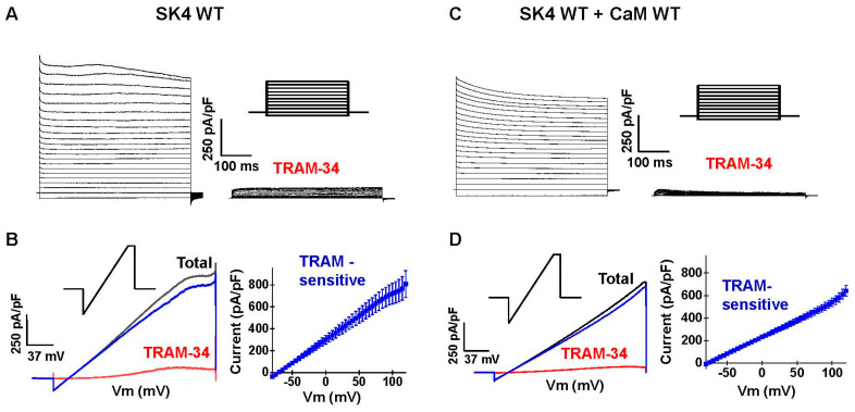
Ca2+ binding to the ubiquitous Ca2+ sensing protein calmodulin (CaM) activates the intermediate conductance Ca2+-activated SK4 channel. Potential hydrophilic pockets for CaM binding have been identified at the intracellular HA and HB helices in the C-terminal of SK4 from the three published cryo-EM structures of SK4. Single charge reversal substitutions at either site, significantly weakened the pull-down of SK4 by CaM wild-type (CaM), and decreased the TRAM-34 sensitive outward K+ current densities in native HEK293T cells when compared with SK4 WT measured under the same conditions. Only the doubly substituted SK4 R352D/R355D (HB helix) obliterated the CaM-mediated pull-down and thwarted outward K+ currents. However, overexpression of CaM E84K/E87K, which had been predicted to face the arginine doublet, restored the CaM-mediated pull-down of SK4 R352D/R355D and normalized its whole-cell current density. Virtual analysis of the putative salt bridges supports a unique role for the positively charged arginine doublet at the HB helix into anchoring the interaction with the negatively charged CaM glutamate 84 and 87 CaM. Our findings underscore the unique contribution of electrostatic interactions in carrying CaM binding onto SK4 and support the role of the C-terminal HB helix to the Ca2+-dependent gating process.

Keywords: calcium; calmodulin; co-immunoprecipitation; electrophysiology; ion channel; protein–protein interaction.


Conflict of interest statement

The authors declare no conflicts of interest with the content of the manuscript. The funders had no role in the design of the study; in the collection, analyses, or interpretation of data; in the writing of the manuscript; or in the decision to publish the results.

Figures

Figure 1
Figure 1
Predicted sets of electrostatic interactions site between CaM and intracellular HA/HB helices of SK4. (A). The cryo-EM structure of the tetrameric SK4 channel in complex with four CaM proteins viewed from the intracellular face. This 3-D model was obtained using the Ca2+-bound 6CNO.pdb structure. The missing extracellular loops were reconstituted by MODELLER9v11. As seen, a single CaM protein interacts with two channel pore-forming α subunits. (B). Details of the intracellular interface between CaM and SK4 viewed from below the cell surface. Only two out of the four pore-forming α subunits are shown for clarity. The region between the S4–S5 linker and the C-terminus was omitted. CaM proteins are depicted in silver whereas the intracellular helices HA and HB for each channel of the two pore-forming α subunit herein depicted are colored in blue and red, respectively. The intracellular HA and HB helices of one channel subunit folds back under the channel in a fashion anti-parallel to the plane of the membrane. The C-lobe of one CaM molecule (CaM-C) is anchored to the HA and HB helices, whereas the N-lobe of the same CaM molecular (CaM-N) forms a complex with the S45A helix of a different pore-forming α subunit. Ca2+ ions are represented as green spheres. (C). Predicted interactions between SK4 HA helix with residues in the CaM C-lobe EF-4. (D). Predicted interactions between the SK4 HB helix and the CaM C-lobe EF-3. Original figures were produced with Discovery Studio.
Figure 2
Figure 2
Robust heterologous expression of SK4 in HEK293T cells. pMT21-hSK4 WT was co-transfected without recombinant (endogenous CaM) or with pIRES-DsRed-HisB-cMyc-CaM WT in HEK293T cells. Whole-cell currents were recorded in the presence of 0.1 µM intracellular free Ca2+ under a [K+ ] gradient of 140 mM (inside)/over 3 mM (out). (A). Representative current traces of SK4 WT recorded from HEK293T cells in the absence (left) and presence (right) of 0.3 µM TRAM-34. Currents were activated by 500 ms step voltages between −90 and +120 mV from a holding potential of −80 mV. The voltage pulse protocol is illustrated above the TRAM-34 current traces. (B). Left. Representative current traces of SK4 WT recorded using a voltage ramp protocol. Cells were depolarized to +120 mV for 50 ms from a holding potential of −80 mV, followed by a 3-s voltage ramp from +120 to −100 mV before (black) and after (red) the application of TRAM-34. The ramp pulse protocol is illustrated above the total current traces. The TRAM-34 sensitive SK4 WT current (blue) was determined by subtracting the ramp current trace obtained after the addition of 0.3 µM TRAM-34 (red) from the total current (black), and the current densities of TRAM-34 sensitive SK4 WT current were plotted as a function of voltage (Right). (C). Representative current traces of SK4 WT co-expressed with CaM WT recorded from HEK293T cells in the absence (left) and presence (right) of 0.3 µM TRAM-34. (D). Left. Representative ramp current traces of SK4 WT + CaM WT before (black) and after (red) the application of TRAM-34. The TRAM-34 sensitive SK4 WT + CaM WT current (blue) was estimated by subtracting the current trace obtained after the addition of TRAM-34 (red) from the total current (black) and plotted as a function of applied voltage (right).
Figure 3
Figure 3
SK4 K327E disrupts SK4/CaM interaction. (A). HEK293T cells were transiently transfected with either pMT21-hSK4 WT or K327E combined with either pIRES-DsRed-HisB-cMyc CaM WT or E127K. (Ai). All proteins were expressed at the expected molecular mass. (Aii). Cell lysates (300 μg) were immunoprecipitated (IP) overnight with anti-His magnetic beads. SK4 WT was pulled down by CaM WT and by CaM E127K. In comparison, the pull-down signal was weaker for SK4 K327E in the presence of CaM WT and CaM E127K (obtained in four different protein lysate preparations during a period of 15 months). (B). HEK293T cells were transiently transfected with either pMT21-hSK4 WT, K327E, combined with either pIRES-DsRed-HisB-cMyc CaM WT, E123K/E127K (K/K) or E123A/E127A (A/A). (Bi). All proteins were expressed at the expected molecular mass. (Bii). Cell lysates (300 μg) were immunoprecipitated (IP) overnight with anti-His magnetic beads. SK4 WT was pulled down by CaM WT, by CaM E123K/E127K, and by CaM E123A/E127A. Pull-down signal was reduced for SK4 K327E + CaM WT but the protein signal was present for the channel pair SK4 K327E + CaM E123K/E127K and the pair SK4 K327E + CaM E123A/E127A confirming that the charge at position Glu123 and Glu127 in CaM may not be a limiting factor in the protein–protein interaction. Similar results were obtained for three different protein lysate preparations during a period of 2 months.
Figure 4
Figure 4
Double substituted channel SK4 R3552D/R355D prevents pull-down by CaM WT. (A). HEK293T cells were transiently transfected with either pMT21-hSK4 WT, R352D (52D), R355D (55D), or R352D/R355D (RD/RD) and pIRES-DsRed-HisB-cMyc CaM WT. Cell lysates (300 μg) were immunoprecipitated (IP) overnight with anti-cHis magnetic beads to capture His-tagged CaM. Immunoblotting was carried out after eluting the protein complexes from the beads with anti-SK4 and anti-His antibodies, the latter identifying recombinant CaM proteins. As seen in the (Ai) panel, total proteins or input fraction, proteins were expressed at the expected molecular mass. (Aii). The protein signal remained strong for SK4 WT after protein elution from the anti-His tagged beads, but signals are considerably weaker for SK4 R352D and near eliminated for SK4 R355D and SK4 R352D/R355D, suggesting the loss of a strong protein interaction. Similar results were obtained for 8 different protein lysate preparations during a period of 36 months. (B). pMT21-hSK4 WT, R352D, R355D or R352D/R355D were transfected in HEK293T cells. Experiments were also carried out in HEK293T cells transfected with the empty pMT21 vector (shown in blue). TRAM-34 sensitive currents were recorded in the presence of 100 nM intracellular free Ca2+ with a [K+] gradient of 140 mM (inside)/3 mM (out) over the −80 mV to +120 mV range. Average current densities are shown ± SEM. Current densities recorded with SK4 R355D (gray) or R352D/R355D (red) were indistinguishable from currents measured in mock-transfected cells (blue). (C). Empty pMT21 vector, pMT21-hSK4 WT, R352D, R355D or R352D/R355D were co-transfected with pIRES-DsRed-HisB-cMyc CaM WT in HEK293T cells. TRAM-34 sensitive currents were recorded under the same conditions as in (C). Overexpression of CaM WT did not improve the SK4 R352D/R355D current density, but significantly increased the current density of SK4 R355D. Average current densities ± SD recorded at +60 mV, including the statistical analysis, are shown in Table 1.
Figure 5
Figure 5
SK4 R352D and SK4 R355D are targeted to the cell surface. HEK293T cells were transiently transfected with SK4 WT and SK4 R352D (Panel A), or SK4 WT and SK4 R355D (Panel B). Two days after transfection, the cells were lysed, and cell fractions were obtained through preparative ultracentrifugation as described in the Experimental procedures section. Western blotting was carried out for the four protein fractions identified as such: lane 1, total proteins; lane 2, cytoplasmic proteins; lane 3, total membrane proteins; and lane 4, enriched plasma membrane proteins. The proteins were probed with the following antibodies: SK4 (Abcam; ab75956, 1:1000 dilution); CaM (Millipore; catalog no.: 05-193, 1:1000 dilution) with anti-mouse (1:10,000 dilution); and cadherin (Pan-cadherin; Thermo Fisher; catalog no.: 71-7100, 1:1000 dilution) with anti-rabbit (1:10,000). Cadherin was used as a marker for the plasma membrane. Each lane was loaded with 20 μg proteins. The molecular markers are shown to the left of the blots with the value provided in kilodalton. The molecular masses were estimated by linear regression and interpolation from the molecular mass markers using the Image Lab software, version 5.2 (Bio-Rad Laboratories (Canada) Ltd, Saint-Laurent, QC H4R 2E).
Figure 6
Figure 6
CaM E84K disrupts the SK4/CaM interaction. (A). HEK293T cells were transiently transfected with pMT21-hSK4 WT, R352D (52D), R355D (55D), or R352D/R355D (RD/RD) and pIRES-DsRed-HisB-cMyc CaM E84K. Cell lysates (300 μg) were immunoprecipitated (IP) overnight with anti-His magnetic beads as described above. (Ai). All proteins were expressed at the expected molecular mass. (Aii). SK4 WT, R355D, and R352D/R355D were not pulled down by CaM E84K but a clear protein signal was observed for R352D. These results indicate that a negatively charged residue at CaM 84 is required to carry constitutive interaction of CaM with SK4 WT and that this interaction could involve a positively charged residue at position 352 in SK4. Similar results were obtained for 3 different protein lysate preparations during a period of 24 months. (B). HEK293T cells were transiently transfected with pMT21-hSK4 WT, R352D (52D), R355D (55D), or R352D/R355D (RD/RD) and pIRES-DsRed-HisB-cMyc CaM WT or E87K. (Bi). All proteins were expressed at the expected molecular mass. (Bii). Cell lysates (300 μg) were immunoprecipitated (IP) overnight with anti-His magnetic beads, as described above. SK4 WT was pulled down by CaM E87K but SK4 R352D, SK4 R355D, and SK4 R352D/R355D were not co-immunoprecipitated. Similar results were obtained for 3 different protein lysate preparations during a period of 24 months.
Figure 7
Figure 7
Charge reversal in both SK4 and CaM rescued interaction and function. (A). HEK293T cells were transiently transfected with pMT21-hSK4 WT, R352D (52D), R355D (55D), or R352D/R355D (RD/RD) and pIRES-DsRed-HisB-cMyc CaM WT or CaM E84K/E87K (EK/EK). (Ai). All proteins were expressed at the expected molecular mass. (Aii). Cell lysates (300 μg) were immunoprecipitated (IP) overnight with anti-His magnetic beads as described above. SK4 WT was not pulled down by CaM E84K/E87K but SK4 R352D and SK4 R352D/R355D were successfully co-immunoprecipitated. Similar results were obtained for 3 different protein lysate preparations during a period of 24 months. (B). TRAM-34 sensitive currents for SK4 WT, R352D, R355D, R352D/R355D, K327E, and R352D/R355D/K327E were measured at 0.1 µM Cai following co-expression with CaM E84K/E87K. Average current densities are shown ± SEM. Complete set of average current densities ± SD recorded at +60 mV, including the statistical analysis, are shown in Table 2. (C). TRAM-34 sensitive currents for SK4 WT, R352D, R355D, R352D/R355D, K327E, and R352D/R355D/K327E were measured at 0.03 µM Cai following co-expression with CaM E84K/E87K. Average current densities are shown ± SEM. Complete set of average current densities ± SD recorded at +60 mV, including the statistical analysis, are shown in Table 2.

Similar articles

References

    1. Meech R. Intracellular calcium injection causes increased potassium conductance in Aplysia. Comp. Biochem. Physiol. A Comp. Physiol. 1972;42:493–499. doi: 10.1016/0300-9629(72)90128-4. - DOI - PubMed
    1. Gardos G. Effect of ethylenediaminetetraacetate on the permeability of human erythrocytes. Acta Physiol. Acad. Sci. Hung. 1958;14:1–5. - PubMed
    1. Klein H., Abu-Arish A., Trinh N.T.N., Luo Y., Wiseman P.W., Hanrahan J.W., Brochiero E., Sauvé R. Investigating CFTR and KCa3.1 Protein/Protein Interactions. PLoS ONE. 2016;11:e0153665. doi: 10.1371/journal.pone.0153665. - DOI - PMC - PubMed
    1. Kshatri A.S., Gonzalez-Hernandez A., Giraldez T. Physiological Roles and Therapeutic Potential of Ca2+ Activated Potassium Channels in the Nervous System. Front. Mol. Neurosci. 2018;11:258. doi: 10.3389/fnmol.2018.00258. - DOI - PMC - PubMed
    1. Skibsbye L., Poulet C., Diness J.G., Bentzen B.H., Yuan L., Kappert U., Matschke K., Wettwer E., Ravens U., Grunnet M., et al. Small-conductance calcium-activated potassium (SK) channels contribute to action potential repolarization in human atria. Cardiovasc. Res. 2014;103:156–167. doi: 10.1093/cvr/cvu121. - DOI - PubMed

MeSH terms

Substances

Grants and funding

LinkOut - more resources