. 2024 Apr 12;25(8):4283.
doi: 10.3390/ijms25084283.

Development of a Novel, Potent, and Selective Sialyltransferase Inhibitor for Suppressing Cancer Metastasis

Affiliations

Development of a Novel, Potent, and Selective Sialyltransferase Inhibitor for Suppressing Cancer Metastasis

Han-En Tsai et al. Int J Mol Sci. .

Abstract

Sialyltransferase-catalyzed membrane protein and lipid glycosylation plays a vital role as one of the most abundant post-translational modifications and diversification reactions in eukaryotes. However, aberrant sialylation has been associated with cancer malignancy and metastasis. Sialyltransferases thus represent emerging targets for the development of small molecule cancer drugs. Herein, we report the inhibitory effects of a recently discovered lithocholic acid derivative FCW393 on sialyltransferase catalytic activity, integrin sialyation, cancer-associated signal transduction, MDA-MB-231 and B16F10 cell migration and invasion, and in in vivo studies, on tumor growth, metastasis, and angiogenesis. FCW393 showed effective and selective inhibition of the sialyltransferases ST6GAL1 (IC50 = 7.8 μM) and ST3GAL3 (IC50 = 9.45 μM) relative to ST3GAL1 (IC50 > 400 μM) and ST8SIA4 (IC50 > 100 μM). FCW393 reduced integrin sialylation in breast cancer and melanoma cells dose-dependently and downregulated proteins associated with the integrin-regulated FAK/paxillin and GEF/Rho/ROCK pathways, and with the VEGF-regulated Akt/NFκB/HIF-1α pathway. FCW393 inhibited cell migration (IC50 = 2.6 μM) and invasion in in vitro experiments, and in in vivo studies of tumor-bearing mice, FCW393 reduced tumor size, angiogenesis, and metastatic potential. Based on its demonstrated selectivity, cell permeability, relatively low cytotoxicity (IC50 = 55 μM), and high efficacy, FCW393 shows promising potential as a small molecule experimental tool compound and a lead for further development of a novel cancer therapeutic.

Keywords: breast cancer; integrin sialylation; melanoma; metastasis; sialyltransferase inhibitor.


Conflict of interest statement

The authors declare no conflicts of interest.

Figures
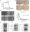
Figure 1
Figure 1
Analysis of cell viability, motility, and Western blot analysis in DMSO (control) and FCW393-treated MDA-MB-231 cells. (A) A plot showing the percentage of viable MDA-MB-231 cells remaining following 48 h incubation with 20–80 μM FCW393. Each data point represents the mean ± SEM of 4 independent measurements. (B) Representative photomicrographs of MDA-MB-231 cells treated with 10 or 30 μM FCW393, or with DMSO and subjected to the wound healing assay. Images were recorded at 0 and 24 h after wound induction; Scale bar: 200 μm. (C) Top panel: Representative photomicrographs of MDA-MB-231 cells treated with 5, 10, or 20 μM FCW393, or DMSO and subjected to the transwell migration assay); Scale bar: 100 μm. Bottom panel: A plot of the percentage of migrated cells vs. [FCW393] showing data fitting to define the IC50. Western blot analysis of integrin/FAK-paxillin/NFκB signal transduction pathway proteins in MDA-MB-231 cells treated with (10 or 20 μM) FCW393, or DMSO (control) for 48 h. β-Tubulin was used as the loading control. The phosphorylated protein forms are represented by prefix “p” and the phosphorylation site is given in parentheses. (D) Western blots of phosphorylated and dephosphorylated talin, intergrins β1–4, and integrins αv and α4. (E) Western blots of FAK, p-FAK, paxillin, and p-paxillin. (F) Western blots of p-IKKα/β, IKKα, IKKβ, IκBα, p-IκBα, NFκB p65, and p-NFκB p65.
Figure 2
Figure 2
Analysis of tumor size and metastasis in nude mice bearing established MDA-MB-231/Luc tumors (100 mm3) and receiving 40% PEG400 + 60% normal saline (control) or FCW393 (10 mg/kg body weight) by intraperitoneal injection every other day. (A). In vivo luciferase bioluminescence images of tumors in mice measured at week 8 of FCW393 treatment. (B). Total tumor bioluminescence vs. treatment period (n = 10 per group). (C). Tumor size vs. treatment period (n = 9; ** p < 0.01). (D). Photomicrographs (100× magnification) of representative H & E-stained lung tumor tissue samples from control and FCW393-treated mice at day 60. The arrows point to the metastatic colonies in control group sample.
Figure 3
Figure 3
Analysis of cell viability, motility, and Western blot analysis in DMSO (control) and FCW393-treated B16F10 cells. (A) A plot showing the percentage of viable B16F10 cells remaining following 4 h incubation with 1–80 μM FCW393. Each data point is the mean ± SEM of 4 independent measurements. (B) Photographs of crystal violet-stained B16F10 cell colonies following incubation with 10, 20, or 30 μM FCW393 for 7 days. (C) Top panel: Representative photomicrographs of traversed B16F10 cells following treatment with 10, 20, or 30 μM FCW393 for 16 h, transmigration through a Matrigel-coated polycarbonate filter (8 μm pore size), fixing, and Giemsa staining); Scale bar: 100 μm. Bottom panel: Percentage of the total number of traversed FCW393 (0, 5, 10, 20, and 30 μM)-treated cells relative to the number of traversed control cells. All data are expressed as mean ± SEM based on 3 independent experiments (** p < 0.01). Western blot analysis of the integrin/GEF/Rho/ROCK and VEGF/Akt/NFκB/HIF-1α signal transduction pathway proteins in B16F10 cells treated with (10, 20, or 30 μM) FCW393 or DMSO (control) for 4 h. β-Tubulin or β-actin were used as the loading control. The phosphorylated protein forms are represented by the prefix “p” and the phosphorylation site is given in parentheses. (D) Western blot of p-FAK, FAK, p-paxillin, and paxillin. (E) Western blot of p-IKKα/β IKKα, IKKβ, IκBα, p-IκBα, NFκB p65, and p-NFκB p65. (F) Western blot of p-VEGFR2, VEGFR2, VEGF, p-Akt, Akt, and HIF-1α. (G) Western blot of Rho A-C, ROCK I & II, and Rac I.
Figure 4
Figure 4
Analysis of tumor size, metastasis, and angiogenesis in C57BL/6JNarl mice bearing established B16F10 cell tumors (100 mm3) and starting on 10 d following implantation, receiving 40% PEG400 + 60% normal saline (control) or FCW393 (10 mg/kg body weight) by intraperitoneal injection every other day. (A) Photographs of FCW393-treated and control mice at day 26 following implantation. Tumors are located within the red circles; * p < 0.05. (B) A plot of (harvested) tumor volume vs. days following implantation and then FCW393 treatment (n = 10 in each group; * p < 0.05; **, p < 0.01). (C) Photomicrographs (10× magnification) of fixed and CD31-immunostained tumor tissue samples at day 26 following initiation of FCW393 treatment (n = 5 in each group); Scale bar: 500 μm. (D) Photomicrographs (20× magnification) of fixed and Ki67-immunostained tumor tissue samples at day 26 following initiation of FCW393 treatment (n = 5 in each group); Scale bar: 200 μm. Determination of the effect of FCW393 on B16F10 cell metastasis. C57BL/6JNarl mice were intravenously administered with luciferase-expressing B16F10 cells at day 0 and starting on day 1, with an intraperitoneal injection of vehicle or FCW393 (10 mg/kg body weight) on alternate days. (E) Bioluminescence images determined at day 7. (F) Bioluminescence images determined at day 14. (G) Total bioluminescence determined for dissected and homogenized lungs. For (EG), data values are reported as the mean ± SEM (n = 6 in each group; * p < 0.05; ** p < 0.01).
Figure 5
Figure 5
Proposed mechanism of FCW393-mediated suppression of tumor growth, inhibition of metastasis, and anti-angiogenesis.

References

    1. Bull C., den Brok M.H., Adema G.J. Sweet escape: Sialic acids in tumor immune evasion. Biochim. Biophys. Acta. 2014;1846:238–246. doi: 10.1016/j.bbcan.2014.07.005. - DOI - PubMed
    1. Reily C., Stewart T.J., Renfrow M.B., Novak J. Glycosylation in health and disease. Nat. Rev. Nephrol. 2019;15:346–366. doi: 10.1038/s41581-019-0129-4. - DOI - PMC - PubMed
    1. Costa A.F., Campos D., Reis C.A., Gomes C. Targeting Glycosylation: A New Road for Cancer Drug Discovery. Trends Cancer. 2020;6:757–766. doi: 10.1016/j.trecan.2020.04.002. - DOI - PubMed
    1. Vajaria B.N., Patel P.S. Glycosylation: A hallmark of cancer? Glycoconj. J. 2017;34:147–156. doi: 10.1007/s10719-016-9755-2. - DOI - PubMed
    1. Garnham R., Scott E., Livermore K.E., Munkley J. ST6GAL1: A key player in cancer. Oncol. Lett. 2019;18:983–989. - PMC - PubMed

LinkOut - more resources