Case Reports
. 2024 Apr 13;25(8):4313.
doi: 10.3390/ijms25084313.

Novel Ultrastructural Insights into the Clear-Cell Carcinoma of the Pancreas: A Case Report

Affiliations
Case Reports

Novel Ultrastructural Insights into the Clear-Cell Carcinoma of the Pancreas: A Case Report

Valentina Giansante et al. Int J Mol Sci. .

Abstract

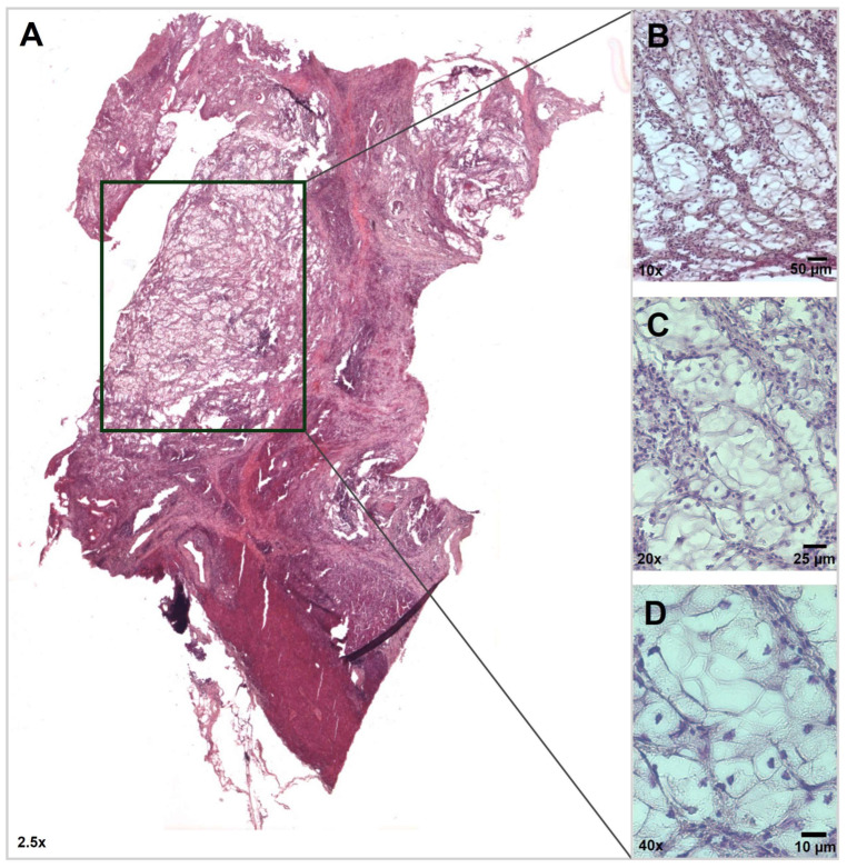
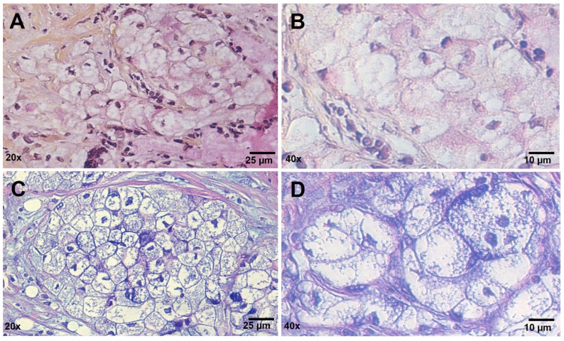
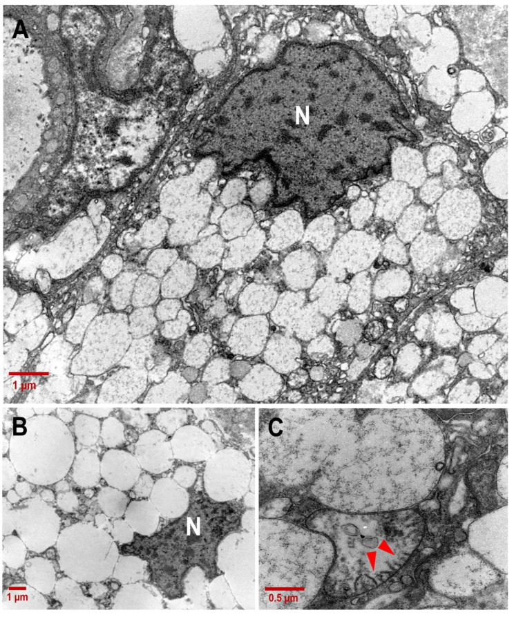
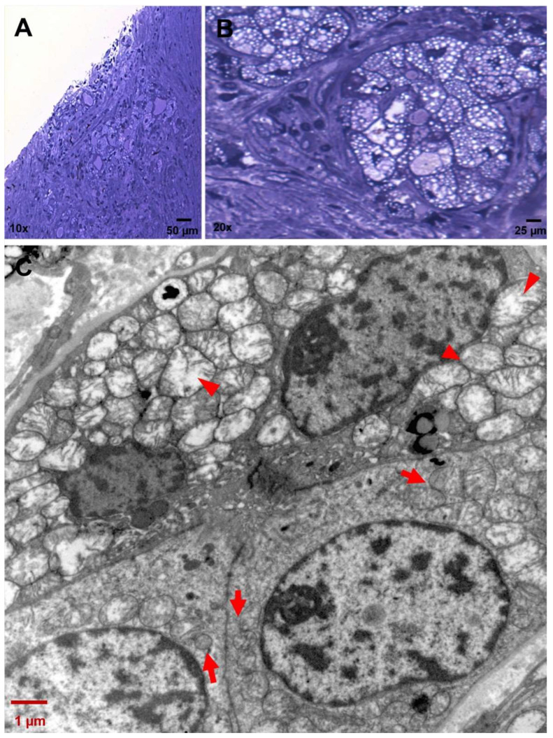
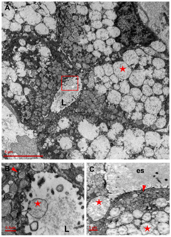
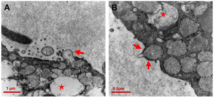
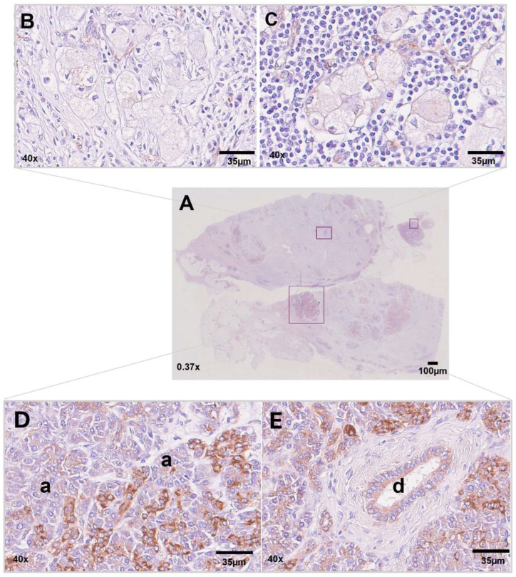
Pancreatic cancer, most frequently as ductal adenocarcinoma (PDAC), is the third leading cause of cancer death. Clear-cell primary adenocarcinoma of the pancreas (CCCP) is a rare, aggressive, still poorly characterized subtype of PDAC. We report here a case of a 65-year-old male presenting with pancreatic neoplasia. A histochemical examination of the tumor showed large cells with clear and abundant intracytoplasmic vacuoles. The clear-cell foamy appearance was not related to the hyperproduction of mucins. Ultrastructural characterization with transmission electron microscopy revealed the massive presence of mitochondria in the clear-cell cytoplasm. The mitochondria showed disordered cristae and various degrees of loss of structural integrity. Immunohistochemistry staining for NADH dehydrogenase [ubiquinone] 1 alpha subcomplex, 4-like 2 (NDUFA4L2) proved specifically negative for the clear-cell tumor. Our ultrastructural and molecular data indicate that the clear-cell nature in CCCP is linked to the accumulation of disrupted mitochondria. We propose that this may impact on the origin and progression of this PDAC subtype.

Keywords: clear-cell primary adenocarcinoma; extracellular vesicles; mitochondria; pancreas; transmission electron microscopy.


Conflict of interest statement

The authors declare no conflicts of interest.

Figures

Figure 1
Figure 1
Histological characterization of the PDAC tissue sample (H&E staining). (A) Whole-slide analysis identifies a prominent area of clear-cell aspect (black box). (BD) At higher magnifications, the tumor shows clear cells with a predominantly solid growth pattern. Scale bars are shown.
Figure 2
Figure 2
CCCP architecture (H&E staining). (A) Nests of clear cells immersed in a desmoplastic stroma (indicated by the black stars) near residual pancreatic acinar epithelial cells. (B) Higher magnification of the clear cells forming nest-like tumor structures. Scale bars are shown.
Figure 3
Figure 3
Mucin histochemical staining. (A,B) Mucicarmine staining; (C,D) Alcian–PAS reaction. Clear cells stained weakly, showing a characteristic thin positive texture within the clear cytoplasm. Scale bars are shown.
Figure 4
Figure 4
Ultrastructure of CCCP cell vacuoles. (A) Clear-cell vacuoles appeared partially electron-lucent under transmission electron microscopy (4400×; scale bar: 1 µm;). (B) Optically empty vacuoles imprinting the nuclear membrane. (C) Structures with the morphology of broken mitochondrial cristae (red arrow heads) were detectable in clear cells (20,000×; scale bar: 0.5 µm). N: nucleus.
Figure 5
Figure 5
Sequential stages of mitochondrial degeneration in CCCP cells. (A,B) Toluidine-blue-stained semithin sections of a CCCP area. Magnification: 10×; 40×. (C) Sequence of mitochondrial involution stages within an acinus: from functional mitochondria with well-preserved cristae (red arrows) to swollen mitochondria with a progressive loss of integrity of cristae (red arrow heads) (4400×; scale bar: 1 µm).
Figure 6
Figure 6
Vacuole distribution in CCCP nests. (A) Clear vacuoles detected both within the cells (red star) and the lumen of a neoplastic acinus (red box) (3000×; scale bar: 5 µm;). (B) Details of an external clear vacuole (red star). The red arrow head shows a cytoplasmic vacuole containing mucin (20,000×; scale bar: 0.5 µm). (C) In the apical portion of a clear cell, small electron-dense subplasmalemmal vacuoles containing mucin (red arrow head) and electron-lucent vacuoles derived by mitochondria (red stars) are easily distinguishable (4400×; scale bar: 1 µm;). L: lumen; es: extracellular space.
Figure 7
Figure 7
Transmembrane progression of vacuoles. (A) An electron-lucent vacuole appears emerging from the plasma membrane (red arrow) (12,000×; scale bar: 1 μm); (B) Protrusions resembling “arms” (red arrows) rise from the membrane to accommodate a mucin-vacuole (20,000×; scale bar 0.5 µm). Disrupted mitochondria are indicated by red stars.
Figure 8
Figure 8
IHC analysis of NDUFA4L2 expression in CCCP cells. (A) Whole-slide scan. (B,C) CCCP showed virtually no NDUFA4L2 expression, with only a weak labeling detected in a few neoplastic cells. (D,E) Strong and diffuse NDUFA4L2 labeling was seen in the non-tumoral areas of the pancreatic tissue. a: acinar epithelial cells and centro-acinar cells; d: excretory duct. Scale bars are shown.

Similar articles

References

    1. Siegel R.L., Miller K.D., Wagle N.S., Jemal A. Cancer statistics, 2023. CA Cancer J. Clin. 2023;73:17–48. doi: 10.3322/caac.21763. - DOI - PubMed
    1. Park W., Chawla A., O’Reilly E.M. Pancreatic Cancer: A Review. JAMA. 2021;326:851–862. doi: 10.1001/jama.2021.13027. - DOI - PMC - PubMed
    1. Krebs A.M., Mitschke J., Lasierra Losada M., Schmalhofer O., Boerries M., Busch H., Boettcher M., Mougiakakos D., Reichardt W., Bronsert P., et al. The EMT-activator Zeb1 is a key factor for cell plasticity and promotes metastasis in pancreatic cancer. Nat. Cell Biol. 2017;19:518–529. doi: 10.1038/ncb3513. - DOI - PubMed
    1. Giansante V., Stati G., Sancilio S., Guerra E., Alberti S., Di Pietro R. The Dual Role of Necroptosis in Pancreatic Ductal Adenocarcinoma. Int. J. Mol. Sci. 2023;24:12633. doi: 10.3390/ijms241612633. - DOI - PMC - PubMed
    1. Truong L.H., Pauklin S. Pancreatic Cancer Microenvironment and Cellular Composition: Current Understandings and Therapeutic Approaches. Cancers. 2021;13:5028. doi: 10.3390/cancers13195028. - DOI - PMC - PubMed

Publication types

MeSH terms