. 2024 Apr 15;25(8):4354.
doi: 10.3390/ijms25084354.

AAV-Mediated CAG-Targeting Selectively Reduces Polyglutamine-Expanded Protein and Attenuates Disease Phenotypes in a Spinocerebellar Ataxia Mouse Model

Affiliations

AAV-Mediated CAG-Targeting Selectively Reduces Polyglutamine-Expanded Protein and Attenuates Disease Phenotypes in a Spinocerebellar Ataxia Mouse Model

Anna Niewiadomska-Cimicka et al. Int J Mol Sci. .

Abstract

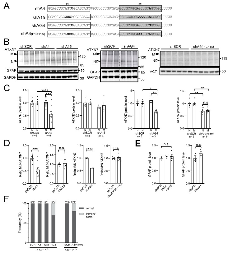
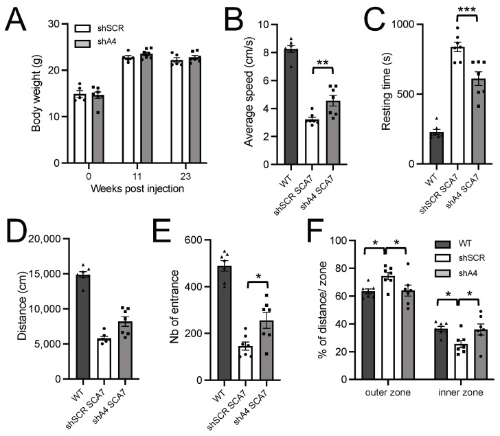
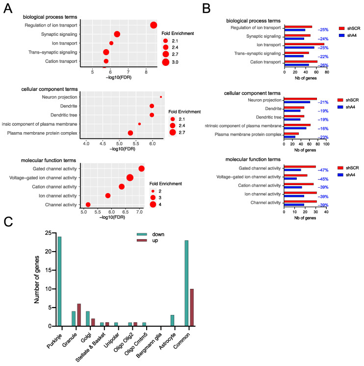
Polyglutamine (polyQ)-encoding CAG repeat expansions represent a common disease-causing mutation responsible for several dominant spinocerebellar ataxias (SCAs). PolyQ-expanded SCA proteins are toxic for cerebellar neurons, with Purkinje cells (PCs) being the most vulnerable. RNA interference (RNAi) reagents targeting transcripts with expanded CAG reduce the level of various mutant SCA proteins in an allele-selective manner in vitro and represent promising universal tools for treating multiple CAG/polyQ SCAs. However, it remains unclear whether the therapeutic targeting of CAG expansion can be achieved in vivo and if it can ameliorate cerebellar functions. Here, using a mouse model of SCA7 expressing a mutant Atxn7 allele with 140 CAGs, we examined the efficacy of short hairpin RNAs (shRNAs) targeting CAG repeats expressed from PHP.eB adeno-associated virus vectors (AAVs), which were introduced into the brain via intravascular injection. We demonstrated that shRNAs carrying various mismatches with the CAG target sequence reduced the level of polyQ-expanded ATXN7 in the cerebellum, albeit with varying degrees of allele selectivity and safety profile. An shRNA named A4 potently reduced the level of polyQ-expanded ATXN7, with no effect on normal ATXN7 levels and no adverse side effects. Furthermore, A4 shRNA treatment improved a range of motor and behavioral parameters 23 weeks after AAV injection and attenuated the disease burden of PCs by preventing the downregulation of several PC-type-specific genes. Our results show the feasibility of the selective targeting of CAG expansion in the cerebellum using a blood-brain barrier-permeable vector to attenuate the disease phenotype in an SCA mouse model. Our study represents a significant advancement in developing CAG-targeting strategies as a potential therapy for SCA7 and possibly other CAG/polyQ SCAs.

Keywords: CAG repeat expansion; CAG-targeting therapy; Spinocerebellar ataxia; adeno-associated virus; allele selectivity; short hairpin RNA.


Conflict of interest statement

The authors declare no competing interests.

Figures

Figure 1
Figure 1
Experimental design: (A) Timeline of disease onset and progression in SCA7140Q/5Q knockin mice. Mice were injected retro-orbitally into their blood stream at an asymptomatic stage of 5–6 weeks of age. A prodromal period is characterized by progressive molecular alterations, notably the accumulation of aggregated mATXN7 observed at 8–10 weeks and the downregulation of PC type-specific genes from 12 weeks. The symptomatic period is characterized by progressive motor and behavioral dysfunctions that manifest from 16 weeks, as revealed by an open field test. The lifespan of SCA7 mice is shortened to about 55–60 weeks. (B) Nucleotide sequence of A2R and scrambled (SCR) shRNAs. Ss, sense strand; as, antisense strand. The as of shA2R corresponds to A2 sd-siRNA [8]. (C) Construct of AAV PHP.eB vector containing two expression cassettes: CMV promoter driving eGFP to trace the efficiency of AAV transduction in vivo and H1 promoter driving shA2R or shSCR. ITR, inverted terminal repeats.
Figure 2
Figure 2
AAV PHP.eB efficiently transduces cerebellar and cerebellar-associated structures. (A) Viral genome copy per cell (vgc/cell) in different mouse tissues 4 days after injection of AAV PHP.eB at increasing doses (vg/kg). Data are expressed as mean ± SEM (n = 4/tissue). (B) Detection of eGFP signal (green) in the majority of PCs on sagittal sections of the cerebellum of mice injected with 1.5 × 10¹³ vg/kg and analyzed 4 weeks post-injection. (C) Higher magnification of PC showing eGFP-positive soma (white arrows) and dendritic arbors (white arrowhead). A number of cells in the molecular layer (yellow arrowheads) (likely stellate and basket neurons) were also eGFP-positive. (DE) An eGFP-positive signal was observed in fibers of striatal neurons (D) and the hippocampal molecular layer (E). (FJ) An eGFP-positive cell soma was detected at high density in the thalamus (F), cerebellar nuclei (G), vestibular and pontine nuclei (H), and the medulla (I), and the spinal cord shows eGFP-positive fibers (J). FN, fastigial nucleus; GCL, granule cell layer; IP, interposed nucleus; IN, intracerebellar nucleus; ML, molecular layer; PC, Purkinje cell; PCL, PC layer. Scale bars: ((B) 1 mm; (C,J) 10 µm; (D) 5 µm; (EI) 50 µm).
Figure 3
Figure 3
Analysis of the efficacy, allele selectivity, and safety of AAV-shA2R injection in SCA7 mice. (A) Representative western blot analysis of soluble normal (N) and mutant (M) ATXN7, Actin-1 (ACT1), and Tyrosine hydroxylase (TH) in the cerebellum of SCA7 mice injected with AAV-shA2R or AAV-shSCR at 1.5 × 10¹³ vg/kg. Mice were injected at the age of 6 weeks and were sacrificed at 10 weeks for analysis. The asterisk indicates 3 mice that developed a tremor-like phenotype. (B) Signal intensities of soluble nATXN7 and mATXN7 were normalized to ACT1 levels and plotted relative to shSCR conditions with the mean set at 1. (C) Ratio of signal intensities of mATXN7/nATXN7 normalized to ACT1 levels. (D) Signal intensities of aggregated mutant ATXN7 in cerebellar protein extracts assessed using on filter trap assay. The intensities were plotted relative to SCR conditions. (E) Dose dependent adverse side effects of shA2R. The affected mice (light grey) either presented with a tremor phenotype and had to be sacrificed or died during the course of the experiment. (F) A 20-nucleotide complementarity to the antisense sequence of shA2R with the mouse Th transcript. (G) Signal intensities of TH normalized to ACT1 levels. Data are expressed as mean± SEM and were analyzed using two-tailed Student’s t-test. * p < 0.05; ** p < 0.01. n.s. for not significant.
Figure 4
Figure 4
In vivo analysis of the efficacy, allele selectivity, and safety of shRNAs A4, A15, AG4, and A4(P10,11). (A) Nucleotide composition of shRNAs. ss, sense strand; as, antisense strand. The as of shA4 and shA15 corresponds to A4 sd-siRNA and A15 sd-siRNA [8]. (B) Representative western blot analysis of ATXN7, ACT1, GFAP, and GAPDH in the cerebellum of SCA7 mice injected with different AAV vectors. The 5-week-old SCA7 mice were injected at 1.5 × 10¹³ vg/kg with AAV-shA4, AAV-shA15, AAV-shAG4, or AAV-shSCR, and at 3.0 × 10¹³ vg/kg with AAV-shA4(P10,11A) and AAV-shSCR. Mice were sacrificed at 5 to 7 weeks post-injection for analysis, depending on the experimental group. Note that the blot in the middle panel was spliced in the center (vertical dotted line) to remove samples from an unrelated treatment and did not have other manipulation. (C) Signal intensities of normal (N) and mutant (M) ATXN7 normalized to GAPDH or ACT1 levels and plotted relative to SCR conditions with mean set at 1. The graph for the experimental group AAV-shA4 and AAV-shSCR combined data from two independently injected cohorts of mice. (D) Ratio of signal intensities of mATXN7/nATXN7 normalized to GAPDH or ACT1 levels and plotted relative to SCR conditions with the mean set at 1. (E) Signal intensities of GFAP normalized to GAPDH levels and plotted relative to SCR conditions with mean set at 1. (F) Frequency of adverse side effects of the different AAV-shRNAs injected at the indicated doses. Data are expressed as mean± SEM and were analyzed using two-tailed Student’s t-test. * p < 0.05; ** p < 0.01; *** p < 0.001; **** p < 0.0001. n.s. for not significant.
Figure 5
Figure 5
Improvement of sensorimotor activity of SCA7 mice treated with AAV-shA4. (A) Post-injection body weight of SCA7 mice treated with AAV-shSCR or AAV-shA4 at 5 weeks of age. Data are mean ± SEM (n = 7 SCA7 males for shA4 and 5 SCA7 males for shSCR). Ordinary two-way ANOVA. (BF) Open field analyses of SCA7 mice treated with AAV-shA4 and AAV-shSCR, and age-matched WT mice. AAV-shA4-treated SCA7 mice had improved locomotor performance and exploration activity at 23 weeks post-injection, compared to control AAV-shSCR-treated SCA7 mice. Data are mean ± SEM. One-way ANOVA followed by post hoc Tukey’s test. * p < 0.05; ** p < 0.01; *** p < 0.001.
Figure 6
Figure 6
Analysis of differentially expressed genes in AAV-shSCR- and AAV-shA4-treated SCA7 mice compared to WT mice. (A) Gene ontology (GO) term enrichment for the 426 DEGs between AAV-shSCR-treated SCA7 mice (n= 5 males) and WT mice (n = 4 males) calculated with ShinyGO v0.80. The 5 most enriched GO terms of each category are sorted by –Log10(FDR). (B) Comparison of the number of DEGs accounting for each major enriched GO term in the transcriptomes of AAV-shSCR- and AAV-shA4-treated SCA7 mice, relative to WT mice. The values in blue represent the percentages of genes (–%) that are deregulated in AAV-shSCR but are restored to a FC < 1.3 or are not deregulated in AAV-shA4-treated SCA7 mice anymore. (C) Cell type-specific distribution of genes that are deregulated in AAV-shSCR but are restored to an FC < 1.3 or are not deregulated in AAV-shA4-treated SCA7 mice anymore. Among the 140 genes deregulated only in AAV-shSCR-treated SCA7 mice, 82 genes were assigned to one or more cerebellar cell types.

Similar articles

Cited by

References

    1. Klockgether T., Mariotti C., Paulson H.L. Spinocerebellar ataxia. Nat. Rev. Dis. Prim. 2019;5:24. doi: 10.1038/s41572-019-0074-3. - DOI - PubMed
    1. Kacher R., Lejeune F.X., Noel S., Cazeneuve C., Brice A., Humbert S., Durr A. Propensity for somatic expansion increases over the course of life in Huntington disease. eLife. 2021;10:e64674. doi: 10.7554/eLife.64674. - DOI - PMC - PubMed
    1. Rub U., Schols L., Paulson H., Auburger G., Kermer P., Jen J.C., Seidel K., Korf H.W., Deller T. Clinical features, neurogenetics and neuropathology of the polyglutamine spinocerebellar ataxias type 1, 2, 3, 6 and 7. Prog. Neurobiol. 2013;104:38–66. doi: 10.1016/j.pneurobio.2013.01.001. - DOI - PubMed
    1. Paulson H.L., Shakkottai V.G., Clark H.B., Orr H.T. Polyglutamine spinocerebellar ataxias—From genes to potential treatments. Nat. Rev. Neurosci. 2017;18:613–626. doi: 10.1038/nrn.2017.92. - DOI - PMC - PubMed
    1. Buijsen R.A.M., Toonen L.J.A., Gardiner S.L., van Roon-Mom W.M.C. Genetics, Mechanisms, and Therapeutic Progress in Polyglutamine Spinocerebellar Ataxias. Neurotherapeutics. 2019;16:263–286. doi: 10.1007/s13311-018-00696-y. - DOI - PMC - PubMed

MeSH terms