Review
. 2024 Apr 16;25(8):4373.
doi: 10.3390/ijms25084373.

Eosinophils in Oral Disease: A Narrative Review

Affiliations
Review

Eosinophils in Oral Disease: A Narrative Review

Huda Moutaz Asmael Al-Azzawi et al. Int J Mol Sci. .

Abstract

The prevalence of diseases characterised by eosinophilia is on the rise, emphasising the importance of understanding the role of eosinophils in these conditions. Eosinophils are a subset of granulocytes that contribute to the body's defence against bacterial, viral, and parasitic infections, but they are also implicated in haemostatic processes, including immunoregulation and allergic reactions. They contain cytoplasmic granules which can be selectively mobilised and secrete specific proteins, including chemokines, cytokines, enzymes, extracellular matrix, and growth factors. There are multiple biological and emerging functions of these specialised immune cells, including cancer surveillance, tissue remodelling and development. Several oral diseases, including oral cancer, are associated with either tissue or blood eosinophilia; however, their exact mechanism of action in the pathogenesis of these diseases remains unclear. This review presents a comprehensive synopsis of the most recent literature for both clinicians and scientists in relation to eosinophils and oral diseases and reveals a significant knowledge gap in this area of research.

Keywords: eosinophils; oral cancer; oral diseases; oral potentially malignant disorders.


Conflict of interest statement

The authors declare no conflicts of interest.

Figures
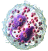
Figure 1
Figure 1
A 3D rendering of eosinophil. From [9].
Figure 2
Figure 2
Schematic representation of our search structure. This diagram is an original creation drawn by the authors.
Figure 3
Figure 3
Clinical features of an oral eosinophilic ulcer on the left border of the tongue extending to the ventral area (Courtesy of A/Prof. Antonio Celentano, Melbourne Dental School, The University of Melbourne, Australia. This photograph is an original creation. All rights reserved).
Figure 4
Figure 4
Lichenoid leukokeratosis of the left retrocommissural area with mild intraepithelial dyskeratosis (courtesy of A/Prof. Antonio Celentano, Melbourne Dental School, The University of Melbourne, Australia. This photograph is an original creation. All rights reserved).
Figure 5
Figure 5
Oral chronic graft versus host disease: widespread lichenoid lesions involving the lower labial mucosa and vermilion border of a patient with acute myeloid leukaemia who underwent allogeneic cell transplant (courtesy of A/Prof. Antonio Celentano, Melbourne Dental School, The University of Melbourne, Australia. This photograph is an original creation. All rights reserved).
Figure 6
Figure 6
Bullous pemphigoid with oral onset, presenting with lesions involving the entire upper alveolar mucosa (courtesy of A/Prof. Antonio Celentano, Melbourne Dental School, The University of Melbourne, Australia. This photograph is an original creation. All rights reserved).
Figure 7
Figure 7
Oral Crohn’s in a paediatric patient presenting with diffuse swelling of the lower third of the face, gross enlargement of the upper and lower lips, and linear ulcers (courtesy of A/Prof. Antonio Celentano, Melbourne Dental School, The University of Melbourne, Australia. This photograph is an original creation. All rights reserved).
Figure 8
Figure 8
Pro-tumourigenic and anti-tumourigenic effects of eosinophils in oral cancer. Pro-tumourigenic activities associated with eosinophils include the activation of gelatinase, which aids in basement membrane degradation and facilitates tumour invasion. Eosinophils also release pre-formed matrix metalloproteinases (MMPs), such as MMP-9 and inhibitory molecules (TIMP-1 and TIMP-2) that participate in extracellular matrix remodelling. Additionally, eosinophils promote tumour angiogenesis by secreting angiogenic factors, including the vascular endothelial growth factor (VEGF), fibroblast growth factor-2 (FGF-2), tumour necrosis factor (TNF), granulocyte-macrophage colony-stimulating factor (GM-CSF), and IL-8. Eosinophils may also contribute to the downregulation of anti-tumour immunity through the secretion of cytokines, such as IL-10 and indoleamine oxidase (IDO). Conversely, eosinophils exhibit anti-tumourigenic effects directly mediated by the killing of tumour cells via eosinophil cationic protein (ECP), major basic protein (MBP), and eosinophil peroxidase (EPO). The release of TNF-a, IL-18, and other cytokines contributes to tumour cytotoxicity either directly or indirectly by stimulating additional effector cells. This figure is an original creation drawn by the authors. All rights reserved.

References

    1. Ehrlich P. Uber die Specifischen Granulation des Blutes. Archiv fur Anatomie und Physiologie. Leipzig, Veit & Company; Leipzig, Germany: 1879. Physiologische Abteilung.
    1. Saraswathi T., Nalinkumar S., Ranganathan K., Umadevi R., Elizabeth J. Eosinophils in health and disease: An overview. J. Oral Maxillofac. Pathol. 2003;7:31–33.
    1. Kovalszki A., Weller P.F. Eosinophilia. Prim. Care. 2016;43:607–617. doi: 10.1016/j.pop.2016.07.010. - DOI - PMC - PubMed
    1. Wardlaw A.J., Moqbel R., Kay A.B. Eosinophils: Biology and role in disease. Adv. Immunol. 1995;60:151–266. - PubMed
    1. Long H., Zhang G., Wang L., Lu Q. Eosinophilic Skin Diseases: A Comprehensive Review. Clin. Rev. Allergy Immunol. 2016;50:189–213. doi: 10.1007/s12016-015-8485-8. - DOI - PubMed

LinkOut - more resources