. 2024 Apr 16;25(8):4374.
doi: 10.3390/ijms25084374.

Effects of Resveratrol on In Vivo Ovarian Cancer Cells Implanted on the Chorioallantoic Membrane (CAM) of a Chicken Embryo Model

Affiliations

Effects of Resveratrol on In Vivo Ovarian Cancer Cells Implanted on the Chorioallantoic Membrane (CAM) of a Chicken Embryo Model

Kenny Chitcholtan et al. Int J Mol Sci. .

Abstract

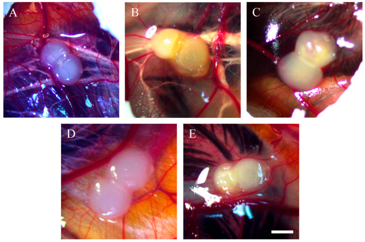
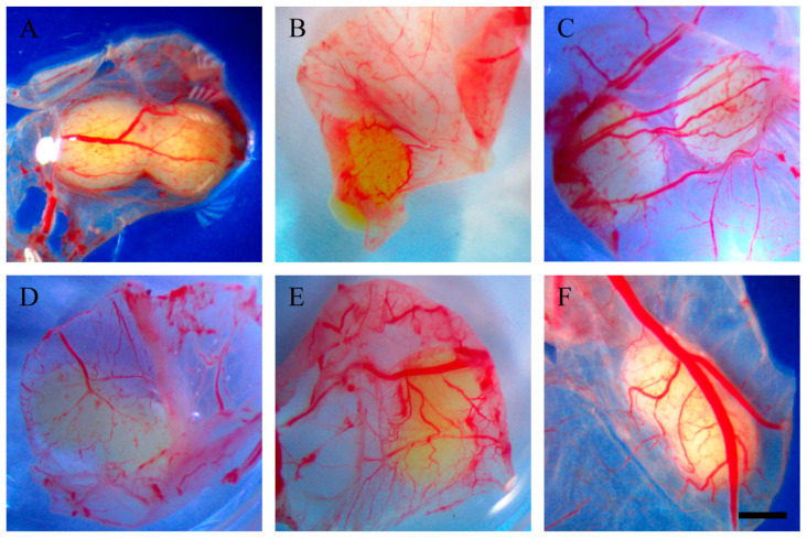
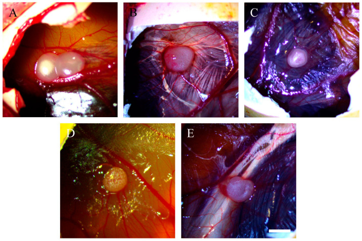
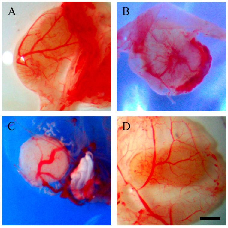
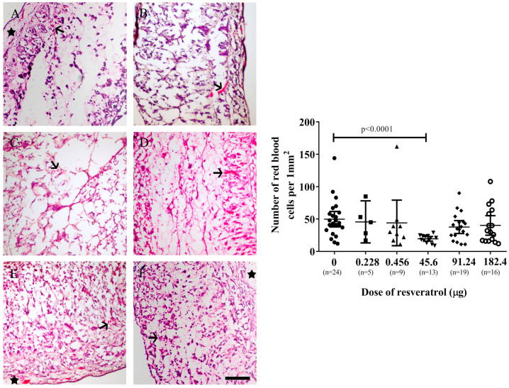
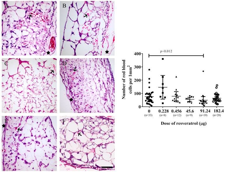
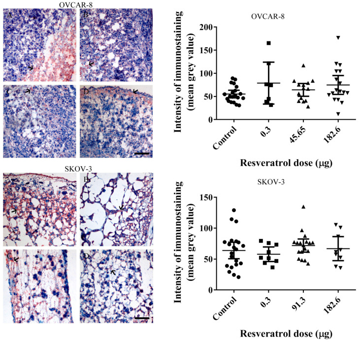
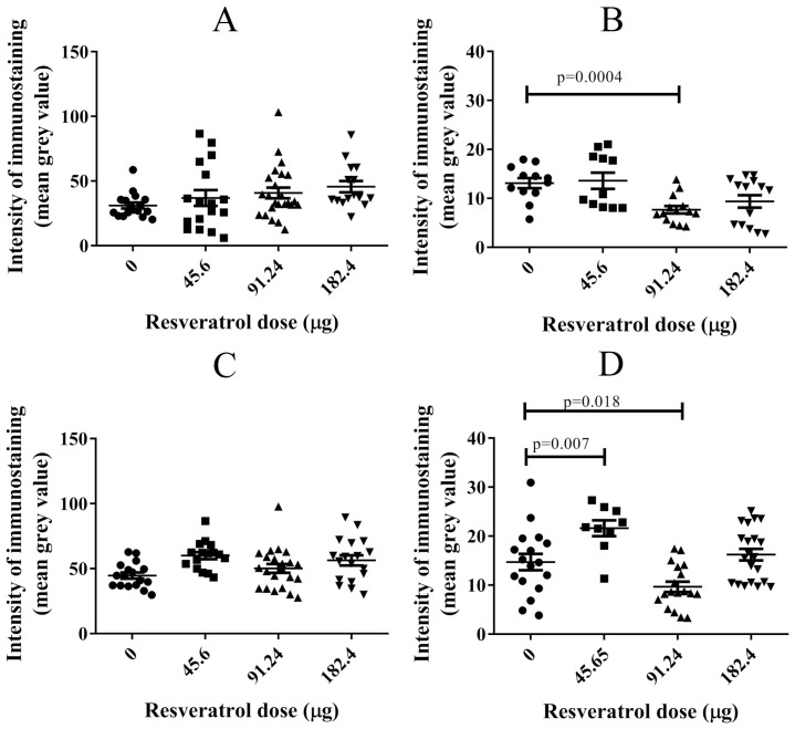
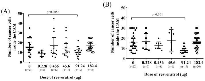
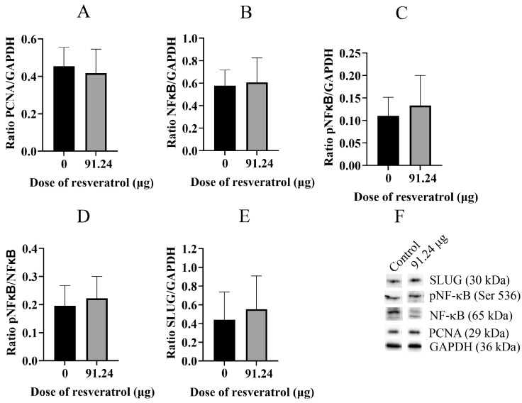
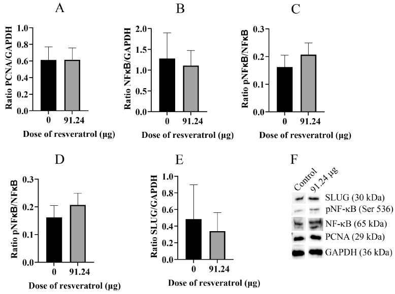
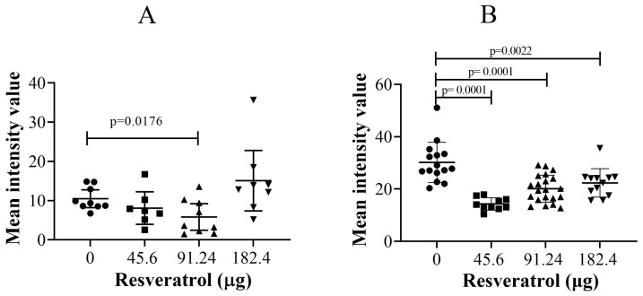
Ovarian cancer poses a significant threat to patients in its advanced stages, often with limited treatment options available. In such cases, palliative management becomes the primary approach to maintaining a reasonable quality of life. Therefore, the administration of any medication that can benefit patients without a curative option holds potential. Resveratrol, a natural compound known for its in vitro anticancer activities, has generated contrasting results in vivo and human studies. In this study, we aimed to assess the anticancer effects of resveratrol on ovarian cancer cells grown on the chorioallantoic membrane (CAM) of chicken embryos. Two ovarian cancer cell lines, OVCAR-8 and SKOV-3, were cultured in collagen scaffolds for four days before being implanted on the CAM of chicken embryos on day 7. Different doses of resveratrol were applied to the CAM every two days for six days. Subsequently, CAM tissues were excised, fixed, and subjected to histological analysis. Some CAM tumours were extracted to analyse proteins through Western blotting. Our findings indicate that specific doses of resveratrol significantly reduce angiogenic activities, pNF-κB levels, and SLUG protein levels by using immunohistochemistry. These results suggest that resveratrol may have the potential to impact the behaviour of ovarian cancer CAM tumours, thereby warranting further consideration as a complementary treatment option for women with incurable ovarian cancer.

Keywords: NF-κB; OVCAR-8; SKOV-3; SLUG; VEGF; cell invasion; chicken embryo; collagen scaffold; ovarian cancer; resveratrol.


Conflict of interest statement

The authors declare no conflicts of interest.

Figures

Figure 1
Figure 1
Stereomicroscopic images of OVCAR-8 tumour implants after 18 days of growth on CAMs. Tumour implants were treated with various doses of resveratrol for six days, starting on day 12 of embryonic development, and the images were captured. Control (A), 0.228 µg (5 µM) (B), 0.456 µg (10 µM) (C), 45.6 µg (1 mM) (D), 91.24 µg (2 mM) (E). Scale bar, 1 mm.
Figure 2
Figure 2
Stereomicroscopic images of excised and inverted OVCAR-8 tumour implants after 18 days of growth on CAMs. Tumour implants were excised from the surrounding CAM and inverted so the vascular networks could be viewed from underneath. Control (A), 0.228 µg (5 µM) (B), 0.456 µg (10 µM) (C), 45.6 µg (1 mM) (D), 91.24 µg (2 mM) (E), 182.4 µg (4 mM) resveratrol (F). Scale bar, 1 mm.
Figure 3
Figure 3
Stereomicroscopic images of SKOV-3 tumour implants after 18 days of growth on CAMs. Tumour implants were with various doses of resveratrol for six days, starting at day 12 of embryonic development, and then images were captured. Control (A), 0.228 µg (5 µM) (B), 0.456 µg (10 µM) (C), 45.6 µg (1 mM) (D), and 91.24 µg (2 mM) resveratrol (E). Scale bar, 1 mm.
Figure 4
Figure 4
Stereomicroscopic images of excised and inverted SKOV-3 tumour implants after 18 days of growth on CAMS. Tumour implants were excised from the surrounding CAM and inverted so the vascular networks could be viewed from underneath. Control (A), 0.228 µg (5 µM) (B), 45.6 µg (1 mM) (C), 91.24 µg (2 mM) resveratrol (D). Scale bar, 1 mm.
Figure 5
Figure 5
Left: Microscopic images of OVCAR-8 tumour implants after six days of treatment with various doses of resveratrol ((A) control, (B) 0.228 µg (5 µM), (C) 0.456 µg (10 µM), (D) 45.6 µg (1 mM), (E) 91.24 µg (2 mM), (F) 182.4 µg (4 mM)). Red blood cells are in red (arrows), and CAM areas are indicated by stars. Right: The number of red blood cells was counted and plotted using various doses of resveratrol. Data are expressed as means ± SEM, n = number of sectioned tumour implants, which were from at least five embryos. Data considered statistically significant compared to controls are indicated as p < 0.05, t-student test. The scale bar is 200 µm.
Figure 6
Figure 6
Left: Microscopic images of SKOV-3 tumour implants after six days of treatment with various doses of resveratrol ((A) control, (B) 0.228 µg (5 µM), (C) 0.456 µg (10 µM), (D) 45.6 µg (1 mM), (E) 91.24 µg (2 mM), (F) 182.4 µg (4 mM)). Red blood cells are in red (arrows), and CAM areas are indicated by stars. Right: The number of red blood cells that were counted and plotted with various doses of resveratrol. Data are expressed as means ± SEM, n = number of sectioned tumour implants, which were from at least five embryos. Data considered statistically significant compared to controls are indicated as p < 0.05, t-student test. The scale bar is 200 µm.
Figure 7
Figure 7
Immunohistological staining of vascular endothelial growth factor (VEGF) in tumour sections. Left-microscopic images of OVCAR-8 (Top: (A) control, (B) 0.3 µg (5 µM), (C) 91.3 µg (2 mM), (D) 182.6 µg (4 mM)) and SKOV-3 (Bottom: (A) control, (B) 0.3 µg (5 µM), (C) 91.3 µg (2 mM), (D) 182.6 µg (4 mM)) tumour implants were immune-stained with an anti-VEGF antibody. The red-brown colour represents levels of VEGF protein (arrows). The intensity of immunostaining of anti-VEGF antibody was subjected to analysis using Fiji ImageJ software 1.53m, and the densitometry of grey value was calculated and plotted. Data are expressed as means ± SEM. The scale bar is 200 µm.
Figure 8
Figure 8
Immunohistological staining of NF-κB (A,C) and pNF-κB (B,D) in OVCAR-8 (A,B) and SKOV-3 (C,D) tumour implants treated with three doses of resveratrol (45.6 (1 mM), 91.24 (2 mM) and 182.4 µg (4 mM)) for six days. Tissue implants were sectioned and stained with anti-NF-κB and phospho-NF-κB antibodies. The histological immunostaining of the antigens was quantitative using Fiji ImageJ software 1.53m. Data are expressed as means ± SEM. Data considered statistically significant compared to controls are indicated as p < 0.05, t-student test.
Figure 9
Figure 9
The number of cancer cells in the area of mesoderm layers of CAM implanted with OVCAR-8 cell line (A) and SKOV-3 (B) treated with various doses of resveratrol. Tumour implants were treated with different doses of resveratrol, 0.228 µg (5 µM), 0.456 µg (10 µM), 45.6 µg (1 mM), 91.24 µg (2 mM), and 182.4 µg (4 mM). Data are expressed as means ± SEM, n = number of sectioned tumour implants, which were from at least five embryos. Data considered statistically significant compared to controls are indicated as p < 0.05, t-student test.
Figure 10
Figure 10
Western blot analysis of specific proteins extracted from whole tumour CAM tissue of the OVCAR-8 cell line. The ratios of protein band intensities for PCNA/GAPDH (A), NFκB/GAPDH (B), pNFκB/GAPDH (C), pNFκB/NFκB (D), and SLUG/GAPDH (E) are presented. Bands of each protein were analysed by Western Blotting (F). A resveratrol dose of 91.24 µg (2 mM) was selected for the inhibition study with tumour implants.
Figure 11
Figure 11
Western blot analysis of specific proteins extracted from whole tumour CAM tissue of the SKOV-3 cell line. The ratios of protein band intensities for PCNA/GAPDH (A), NFκB/GAPDH (B), pNFκB/GAPDH (C), pNFκB/NFκB (D), and SLUG/GAPDH (E) are presented. Bands of each proteins were analysed by Western Blotting (F). A resveratrol dose of 91.24 µg (2 mM) was selected for the inhibition study with tumour implants.
Figure 12
Figure 12
Immunohistochemical staining of SLUG antigens in OVCAR-8 (A) and SKOV-3 (B) tumour implants treated with three doses of resveratrol (45.6 (1 mM), 91.24 (2 mM), and 182.4 (4 mM) µg) for six days. The immunostaining of the antigens was quantitated using Fiji ImageJ software 1.53m. Data considered statistically significant compared to controls are indicated as p < 0.05, t-student test.

Similar articles

Cited by

References

    1. Mukherjee S., Dudley J.I., Das D.K. Dose-dependency of resveratrol in providing health benefits. Former. Nonlinearity Biol. Toxicol. Med. 2010;8:478–500. doi: 10.2203/dose-response.09-015.Mukherjee. - DOI - PMC - PubMed
    1. Raj M.H.G., Abd Elmageed Z.Y., Zhou J., Gaur R.L., Nguyen L., Azam G.A., Braley P., Rao P.N., Fathi I.M., Ouhtit A. Synergistic action of dietary phyto-antioxidants on survival and proliferation of ovarian cancer cells. Gynecol. Oncol. 2008;110:432–438. doi: 10.1016/j.ygyno.2008.05.001. - DOI - PMC - PubMed
    1. Jeonga K.J., Choa K.H., Panupinthub N., Kimc H., Kanga J., Parka C.G., Millsb G.B., Leea H.Y. EGFR mediates LPA-induced proteolytic enzyme expression and ovarian cancer invasion: Inhibition by resveratrol. Mol. Oncol. 2013;7:121–129. doi: 10.1016/j.molonc.2012.10.001. - DOI - PMC - PubMed
    1. Park S.Y., Jeong K.J., Lee J., Yoon D.S., Choi W.S., Kim Y.K., Han J.W., Kim Y.M., Kim B.K., Lee H.Y. Hypoxia enhances LPA-induced HIF-1a and VEGF expression: Their inhibition by resveratrol. Cancer Lett. 2007;258:63–69. doi: 10.1016/j.canlet.2007.08.011. - DOI - PubMed
    1. Vergara D., Simeone P., Toraldo D., Boccio P.D., Vergaro V., Leporatti S., Pieragostino D., Tinelli A., De Domenico S., Alberti S., et al. Resveratrol downregulates Akt/GSK and ERK signalling pathways in OVCAR-3 ovarian cancer cells. Mol. BioSystems. 2012;8:1078–1087. doi: 10.1039/c2mb05486h. - DOI - PubMed

MeSH terms