. 2024 Apr 17;25(8):4404.
doi: 10.3390/ijms25084404.

Regulation of Mertk Surface Expression via ADAM17 and γ-Secretase Proteolytic Processing

Affiliations

Regulation of Mertk Surface Expression via ADAM17 and γ-Secretase Proteolytic Processing

Kevin C Lahey et al. Int J Mol Sci. .

Abstract

Mertk, a type I receptor tyrosine kinase and member of the TAM family of receptors, has important functions in promoting efferocytosis and resolving inflammation under physiological conditions. In recent years, Mertk has also been linked to pathophysiological roles in cancer, whereby, in several cancer types, including solid cancers and leukemia/lymphomas. Mertk contributes to oncogenic features of proliferation and cell survival as an oncogenic tyrosine kinase. In addition, Mertk expressed on macrophages, including tumor-associated macrophages, promotes immune evasion in cancer and is suggested to act akin to a myeloid checkpoint inhibitor that skews macrophages towards inhibitory phenotypes that suppress host T-cell anti-tumor immunity. In the present study, to better understand the post-translational regulation mechanisms controlling Mertk expression in monocytes/macrophages, we used a PMA-differentiated THP-1 cell model to interrogate the regulation of Mertk expression and developed a novel Mertk reporter cell line to study the intracellular trafficking of Mertk. We show that PMA treatment potently up-regulates Mertk as well as components of the ectodomain proteolytic processing platform ADAM17, whereas PMA differentially regulates the canonical Mertk ligands Gas6 and Pros1 (Gas6 is down-regulated and Pros1 is up-regulated). Under non-stimulated homeostatic conditions, Mertk in PMA-differentiated THP1 cells shows active constitutive proteolytic cleavage by the sequential activities of ADAM17 and the Presenilin/γ-secretase complex, indicating that Mertk is cleaved homeostatically by the combined sequential action of ADAM17 and γ-secretase, after which the cleaved intracellular fragment of Mertk is degraded in a proteasome-dependent mechanism. Using chimeric Flag-Mertk-EGFP-Myc reporter receptors, we confirm that inhibitors of γ-secretase and MG132, which inhibits the 26S proteasome, stabilize the intracellular fragment of Mertk without evidence of nuclear translocation. Finally, the treatment of cells with active γ-carboxylated Gas6, but not inactive Warfarin-treated non-γ-carboxylated Gas6, regulates a distinct proteolytic itinerary-involved receptor clearance and lysosomal proteolysis. Together, these results indicate that pleotropic and complex proteolytic activities regulate Mertk ectodomain cleavage as a homeostatic negative regulatory event to safeguard against the overactivation of Mertk.

Keywords: ADAM-17 ectodomain shedding; Gas6; Mertk; intracellular receptor trafficking; membrane expression; proteolysis; γ-secretase.


Conflict of interest statement

The authors declare no conflicts of interest. The funders had no role in the design of the study, in the collection, analyses, or interpretation of data, in the writing of the manuscript, or in the decision to publish the results.

Figures

Figure 1
Figure 1
Differentiation of THP-1 cells induced by PMA. (A) THP-1 monocytes are differentiated to macrophages and induced to express Mertk by the addition of 100 ng/mL of PMA for at least 24 h, while Axl expression is not induced. Likewise, Tyro3 expression remains stable and is unaffected by treatment. H1299 and Beas2B cells are used as positive controls for Mertk/Axl and Tyro3, respectively. The observed doublet for Mertk corresponds to fully glycosylated and partially glycosylated glycoforms. (B) THP-1 cells treated with 100 ng/mL of PMA over 48 h exhibit a decreased expression of Gas6 but an increased expression of Protein S. (C) MertK and Gas6 expression patterns over a time of 48 h after 100 ng/mL of PMA treatment as quantified from Western blots. (D) Bar plots showing MerTK and Gas6 expression quantified from Western blots at 0 h and post 72 h of 100 ng/mL of PMA treatment (significant differences were observed between the 0 h and 72 h time points for Gas6 (* p = 0.029) and MerTK (** p = 0.003), as determined by independent t-tests). (E) THP-1 cells were treated with 100 ng/mL of PMA over 72 h. mRNA was analyzed via qRT-PCR for Mertk, Axl, Protein S (ProS), Tyro3, and Gas6. Mertk transcription was highly elevated, more than 10-fold, due to PMA treatment, while Gas6 transcription was repressed more than 10-fold. (F) PMA treatment of THP-1s illustrates a complimentary phenomenon. As a monocyte, Gas6 is expressed with little Mertk expression; however, when differentiated, Mertk is highly expressed in favor of Gas6.
Figure 2
Figure 2
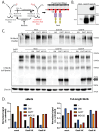
Mertk is sequentially cleaved by ADAM17 and γ-secretase. (A) PMA-differentiated THP-1s (100 ng/mL) express Mertk and the necessary cleavage components, ADAM17 and γ-secretase (nicastrin, PEN2, Pres1, and Pres2; APH-1 not shown) Mertk expression confirmed by both N-terminal-specific (N-Mertk) and C-terminal-specific (C-Mertk) antibodies. Note the expression of an unidentified 90 kDa band. An increase in the pro-form (130 kDa) of ADAM17 is observed within 6 h of PMA treatment, though the mature form (100 kDa) remains constant. (B) PMA-differentiated THP-1s (for 72 h) were treated for 4 h with either 3 µM of GW280264X (GW; ADAM17 inhibitor), 5 µM of DAPT (γ-secretase inhibitor), or 10 µM of MG132 (proteasomal inhibitor). Both DAPT and MG132 treatment results in a band at approximately 50 kDa, with the MG132 band being slightly lower, suggesting the stabilization of mb-cMertk and cyto-cMertk. The slight difference in molecular weight is attributed to the loss of the transmembrane domain. The addition of GW280264X decreases the presence of both fragments due to the sequential nature of Mertk cleavage. In addition, GW280264X slightly increases full-length Mertk expression, demonstrating the enhanced stability of Mertk. Note the presence of the unidentified 90 kDa band. (C) THP-1 cells differentiated with 100 ng/mL of PMA for 72 h were treated with 5 µM of DAPT over the course of 4 h in serum-free media. Stabilization and the subsequent accumulation of the mb-cMertk fragment are observed robustly after 4 h. Lane 5 represents differentiated THP-1 cells after 4 h in untreated serum-free media. (D) Mertk is a substrate of ADAM17 and γ-secretase cleavage. Cleavage occurs sequentially. 1. The ADAM17 cleavage site is identified. 2. Active ADAM17 cleaves Mertk, releasing a soluble ectodomain (sMertk) and leaving a membrane-bound C-terminal fragment (mb-cMertk). 3. After ADAM17 cleavage, the γ-secretase complex identifies the remaining membrane-bound fragment of Mertk. 4. γ-secretase cleavage is initiated and releases a C-terminal fragment into the cytosol (cyto-cMertk).
Figure 3
Figure 3
γ-carboxylated Gas6 Reduces Full-length Mertk Expression and Decreases sMertk. (A) γ-carboxylation status (Gla) of Gas6 is regulated with the addition of Vitamin K or warfarin, producing either a γ-carboxylated, active ligand or a non-γ-carboxylated, inactive ligand, respectively. The carboxylation status (Gla) domain of Gas6 can bind with externalized PS. (B) Recombinant inactive (Gas6-W) and active (Gas6-VK) were produced via HEK293 transfection, with a mock transfection control (Mock). The amount of Gas6 was observed (bottom), while the carboxylation status that is responsible for ligand activity was determined for each (left, top). (C) PMA-differentiated THP-1s were treated for 4 h with either serum-free RPMI only (-), a mock transfection control, 10 nM of active Gas6 (Gas6-VK), or 10 nM of inactive Gas6 (Gas6-W). Treatments were alone or combined with inhibitors of 3 µM of GW280264X (an ADAM17 inhibitor), 5 µM of DAPT (a γ-secretase inhibitor), or 10 µM of MG132 (a proteasomal inhibitor). (D) Quantitative results indicate that Gas6, either active or inactive, does not stabilize the C-terminal fragments as shown in the DAPT and MG132 treatments (bands at 75 kDa). As denoted by sMertk (top), the cleavage is decreased with GW280264X treatment compared to the untreated control.
Figure 4
Figure 4
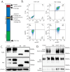
Expression and Validation of tagged Mertk construct in THP-1 cells. (A) A diagram of the tagged Mertk construct is shown to scale to illustrate the size of the FLAG, EmGFP, and Myc tags compared to the Mertk receptor. These tags were added to track both the N-terminal and C-terminal domains post-cleavage. Shown are the IFNγR2 signal peptide (red), FLAG-tag (orange), extracellular N-terminal Mertk region (blue), ADAM17 cleavage site (yellow), transmembrane domain (pink), γ-secretase cleavage site (purple), intracellular C-terminal Mertk region (purple), EmGFP tag (green), and Myc tag (orange). (B) The expression of the tagged construct was assessed via flow cytometry using anti-FLAG-PE (left) and anti-N-Mertk-APC (right). Both signals positively correlate with the intracellular EmGFP signal. (C) Expression of the tagged construct was assessed via Western blotting. Cells were treated for 24 h with either tunicamycin (TM) or Brefeldin A (BFA). The untreated (UT) sample was kept in serum-free RPMI media for the duration of the treatment. TM prevents complete glycosylation and results in a 136 kDa band corresponding to the theoretical molecular weight of the construct, while BFA only partially inhibits glycosylation. (D) The cleavage of the tagged construct is identical to the native Mertk (from Figure 2).
Figure 5
Figure 5
γ-carboxylated Gas6 reduces the tagged Mertk construct on cell membranes of THP-1 cells. (A) THP-1s expressing the tagged construct were starved for 18 h in serum-free RPMI and treated for 3 h. Flow cytometry data shows positive GFPs without staining. As expected, CHX treatment after 3 h reduces the expression of the construct. Gas6-VK, used at a concentration > 10 nM, decreases the FLAG-PE signal more when compared to other treatments known to induce cleavage (PMA, LPS). GW280264X is used as a control for Mertk cleavage. (B) Histogram analysis of “A.” Gas6-VK induces a shift in the FLAG-PE signal, showing that less surface Mertk is present. γ-Carboxylated Gas6 induced the degradation of Mertk on the cell membrane. (C) THP-1s treated with γ-Carboxylated Gas6 at a 10 nM concentration show increased MerTK phosphorylation, detected by immunoblotting against pMerTK. (D) THP-1s expressing the tagged construct, treated with >10 nM of Gas6-VK + 1 µM of PS, show a reduced level of both domains of the tagged construct, suggesting that the Gas6-VK + PS treatment is leading to a degradation of the full-length receptor.
Figure 6
Figure 6
Confocal imaging of the GFP-tagged MerTK construct displayed the differential localization of MerTK upon ligand stimulation and the inhibition of proteases. (A) DAPT and MG132 treatments induce increased cytoplasmic GFP signals compared to untreated cells. (B) Confocal imaging shows GFP localized on the cell membrane, indicating the presence of tagged Mertk constructs on the cell surface. γ-Carboxylated Gas6-treated tagged THP-1 cells showed a reduction in the MerTK construct from the membrane and the localization in lysosomes. (C) Quantification of the GFP fluorescence intensity per cell, calculated from confocal images in mock, γ-carboxylated Gas6, and non-γ-carboxylated Gas6, with the bar plots showing the mean and standard error of each treatment. ns; non-significant; *** p < 0.001; **** p < 0.0001.
Figure 7
Figure 7
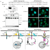
γ-carboxylation of Gas6 induces Mertk degradation independent of phosphorylation. (A) Mutants of the tagged construct were created. K619M, a substitution in the ATP-binding site (underlined) of the Mertk kinase domain, inhibits autophosphorylation by preventing ATP binding and the exchange of phosphate molecules. * Amino acid position for ADAM17 cleavage. (B) Constructs are treated with 10 nM ofGas6-VK for 30 min after a 6 h serum starvation. With the ability to bind ATP and phosphorylate, the WT construct becomes phosphorylated while the kinase dead K619M mutant does not. (C) Mutant constructs were treated with 3 µM of GW280264X (an ADAM17 inhibitor; GW), 5 µM of DAPT (a γ-secretase inhibitor), or 10 µM of MG132 (a proteasomal inhibitor) for 4 h. Results show that the absence of the K619M C-terminal fragment from the MG132 treatment with the addition of GW indicates the fragment is a product of cleavage. (D) Confocal images indicated that the K619M mutants have more cytoplasmatic c-Mertk than WT Mertk with MG132 treatment. (E) Comparison of the contrasting pathways between Notch Receptor and MerTK Signaling Models. In the absence of a ligand, Notch is not cleaved, while MerTK undergoes homeostatic cleavage, leading to proteasomal degradation. Upon ligand binding, Notch is cleaved at the ADAM 17 site, revealing the gamma-secretase site. Subsequent gamma-secretase cleavage releases the Notch intracellular domain, translocating it to the nucleus for transcriptional activation. Conversely, MerTK, upon ligand (Gas6) interaction, is internalized into endosomal compartments and localizes within lysosomes.

Similar articles

Cited by

References

    1. Lemke G. Biology of the TAM receptors. Cold Spring Harb. Perspect. Biol. 2013;5:a009076. doi: 10.1101/cshperspect.a009076. - DOI - PMC - PubMed
    1. Lemke G., Rothlin C.V. Immunobiology of the TAM receptors. Nat. Rev. Immunol. 2008;8:327–336. doi: 10.1038/nri2303. - DOI - PMC - PubMed
    1. Rothlin C.V., Ghosh S., Zuniga E.I., Oldstone M.B., Lemke G. TAM receptors are pleiotropic inhibitors of the innate immune response. Cell. 2007;131:1124–1136. doi: 10.1016/j.cell.2007.10.034. - DOI - PubMed
    1. Lemke G. How macrophages deal with death. Nat. Rev. Immunol. 2019;19:539–549. doi: 10.1038/s41577-019-0167-y. - DOI - PMC - PubMed
    1. McShane L., Tabas I., Lemke G., Kurowska-Stolarska M., Maffia P. TAM receptors in cardiovascular disease. Cardiovasc. Res. 2019;115:1286–1295. doi: 10.1093/cvr/cvz100. - DOI - PMC - PubMed

MeSH terms

LinkOut - more resources