

Brain Abnormalities in Schizophrenia: A Comparative Imagistic Study
- PMID: 38674210
- PMCID: PMC11052149
- DOI: 10.3390/medicina60040564
Brain Abnormalities in Schizophrenia: A Comparative Imagistic Study
Abstract
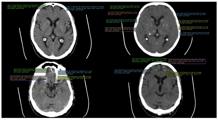
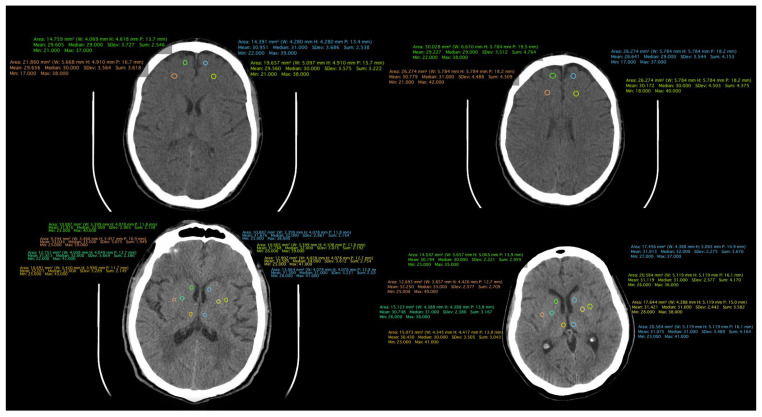
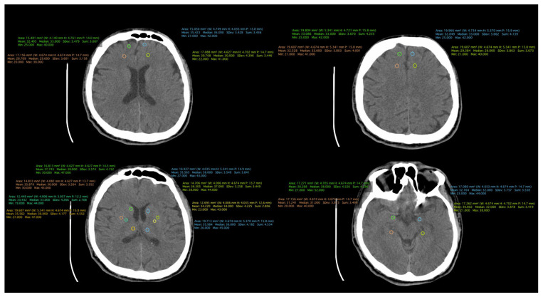
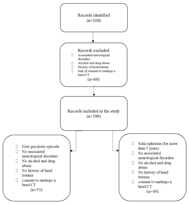
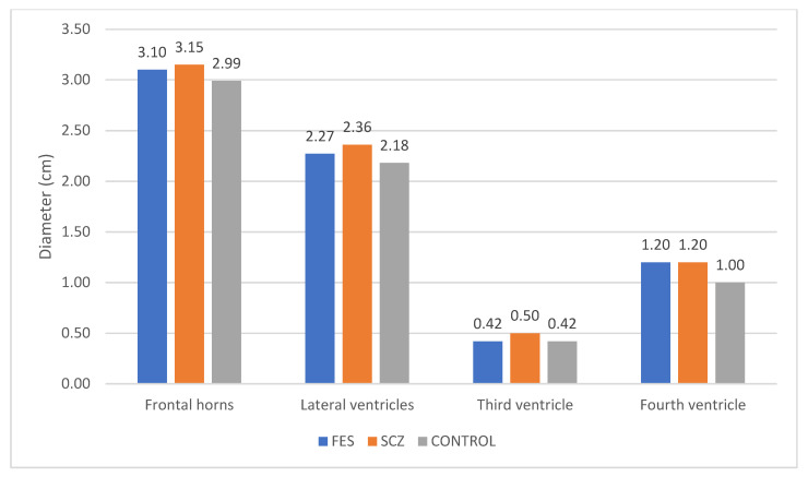
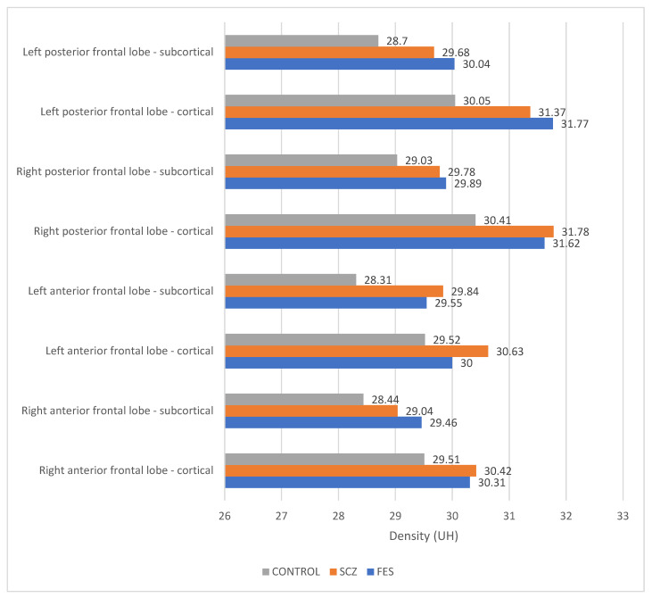
Background and Objectives: Neuroimaging reveals a link between psychiatric conditions and brain structural-functional changes, prompting a paradigm shift in viewing schizophrenia as a neurodevelopmental disorder. This study aims to identify and compare structural brain changes found during the first schizophrenia episode with those found after more than 5 years of illness. Materials and Methods: This prospective study involved 149 participants enrolled between 1 January 2019 and 31 December 2021. The participants were categorized into three groups: the first comprises 51 individuals with an initial psychotic episode, the second consists of 49 patients diagnosed with schizophrenia for over 5 years, and a control group comprising 50 individuals without a diagnosis of schizophrenia or any other psychotic disorder. All participants underwent brain CT examinations. Results: The study examined all three groups: first-episode schizophrenia (FES), schizophrenia (SCZ), and the control group. The FES group had a mean age of 26.35 years and a mean duration of illness of 1.2 years. The SCZ group, with a mean age of 40.08 years, had been diagnosed with schizophrenia for an average of 15.12 years. The control group, with a mean age of 34.60 years, had no schizophrenia diagnosis. Structural measurements revealed widening of frontal horns and lateral ventricles in the SCZ group compared to FES and the FES group compared to the control group. Differences in the dimensions of the third ventricle were noted between SCZ and FES, while no distinction was observed between FES and the control group. The fourth ventricle had similar measurements in FES and SCZ groups, both exceeding those of the control group. Our results showed higher densities in the frontal lobe in schizophrenia patients compared to FES and the control group, with the control group consistently displaying the lowest densities. Conclusions: In summary, our comparative imaging analysis of schizophrenia patients, first-episode schizophrenia, and control patients revealed distinct ventricular patterns, with SCZ showing greater widening than FES and FES wider than the control group. Frontal lobe density, assessed via cerebral CT scans, indicated a higher density in the SCZ group in both anterior and posterior cortex portions compared to FES and the control group, while the left posterior cortex in FES had the highest density. These findings highlight unique neuroanatomical features across groups, shedding light on structural differences associated with different stages of schizophrenia.
Keywords: cerebral CT (computed tomography); cerebral density; first psychotic episode; neuroimaging; schizophrenia.
Conflict of interest statement
The authors have no conflicts of interest to declare.
Figures
Similar articles
-
Abnormalities of regional homogeneity and its correlation with clinical symptoms in Naïve patients with first-episode schizophrenia.Brain Imaging Behav. 2019 Apr;13(2):503-513. doi: 10.1007/s11682-018-9882-4. Brain Imaging Behav. 2019. PMID: 29736883
-
Structural and functional brain abnormalities in schizophrenia: A cross-sectional study at different stages of the disease.Prog Neuropsychopharmacol Biol Psychiatry. 2018 Apr 20;83:27-32. doi: 10.1016/j.pnpbp.2017.12.017. Epub 2017 Dec 29. Prog Neuropsychopharmacol Biol Psychiatry. 2018. PMID: 29292241
-
Brain anatomical abnormalities in high-risk individuals, first-episode, and chronic schizophrenia: an activation likelihood estimation meta-analysis of illness progression.Schizophr Bull. 2011 Jan;37(1):177-88. doi: 10.1093/schbul/sbp073. Epub 2009 Jul 24. Schizophr Bull. 2011. PMID: 19633214 Free PMC article.
-
Morphometric Analysis of Structural MRI Using Schizophrenia Meta-analytic Priors Distinguish Patients from Controls in Two Independent Samples and in a Sample of Individuals With High Polygenic Risk.Schizophr Bull. 2022 Mar 1;48(2):524-532. doi: 10.1093/schbul/sbab125. Schizophr Bull. 2022. PMID: 34662406 Free PMC article.
-
Neural Activity Alterations and Their Association With Neurotransmitter and Genetic Profiles in Schizophrenia: Evidence From Clinical Patients and Unaffected Relatives.CNS Neurosci Ther. 2025 Feb;31(2):e70218. doi: 10.1111/cns.70218. CNS Neurosci Ther. 2025. PMID: 39924342 Free PMC article. Review.
Cited by
-
Schizophrenia diagnosis based on diverse epoch size resting-state EEG using machine learning.PeerJ Comput Sci. 2024 Aug 20;10:e2170. doi: 10.7717/peerj-cs.2170. eCollection 2024. PeerJ Comput Sci. 2024. PMID: 39314693 Free PMC article.
-
Cerebral Computed Tomographic Findings in Schizophrenia: Relationship to Second-Generation Antipsychotics and Hyperprolactinemia.Healthcare (Basel). 2024 Jul 5;12(13):1343. doi: 10.3390/healthcare12131343. Healthcare (Basel). 2024. PMID: 38998877 Free PMC article.
References
-
- Rozycki M., Satterthwaite T.D., Koutsouleris N., Erus G., Doshi J., Wolf D.H., Fan Y., Gur R.E., Gur R.C., Meisenzahl E.M., et al. Multisite Machine Learning Analysis Provides a Robust Structural Imaging Signature of Schizophrenia Detectable across Diverse Patient Populations and within Individuals. Schizophr. Bull. 2018;44:1035–1044. doi: 10.1093/schbul/sbx137. - DOI - PMC - PubMed
Publication types
MeSH terms
LinkOut - more resources
Full Text Sources
Medical
Miscellaneous
