

Uterine Cesarean Scar Tissue-An Immunohistochemical Study
- PMID: 38674297
- PMCID: PMC11051969
- DOI: 10.3390/medicina60040651
Uterine Cesarean Scar Tissue-An Immunohistochemical Study
Abstract
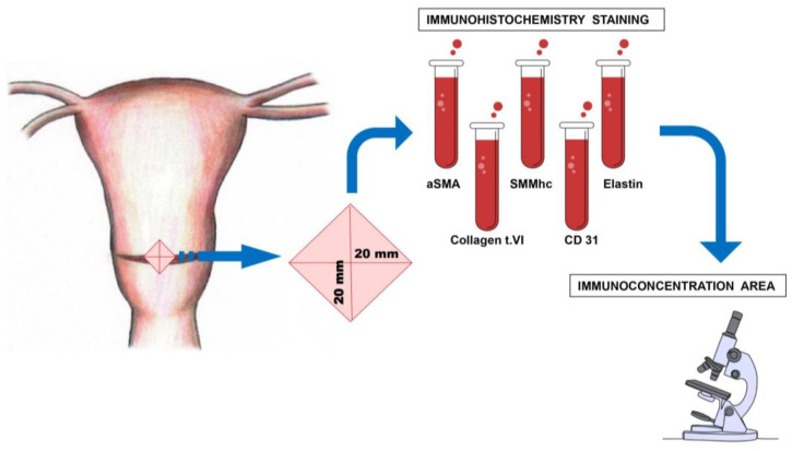
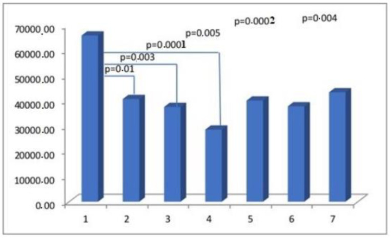
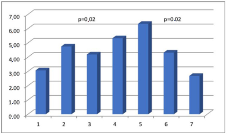
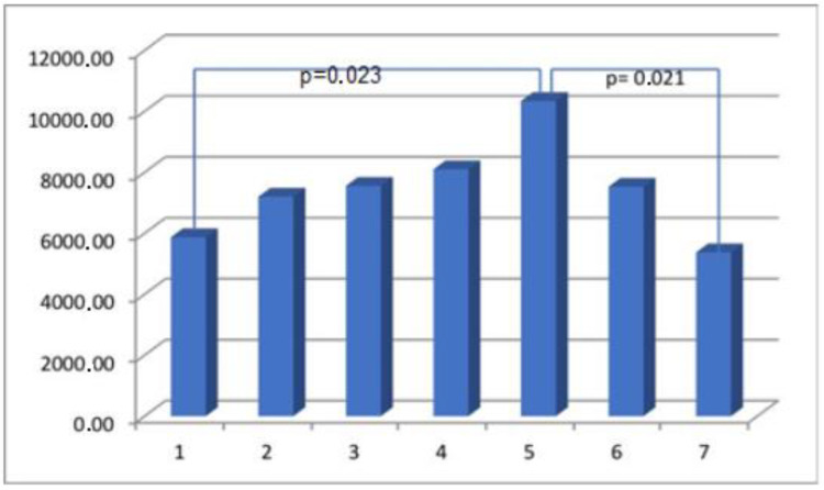
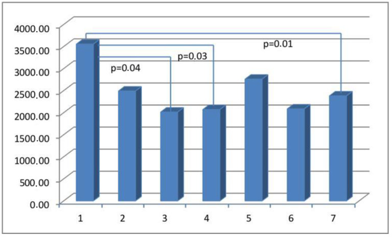
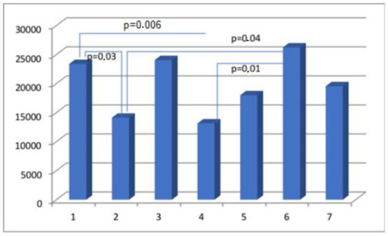
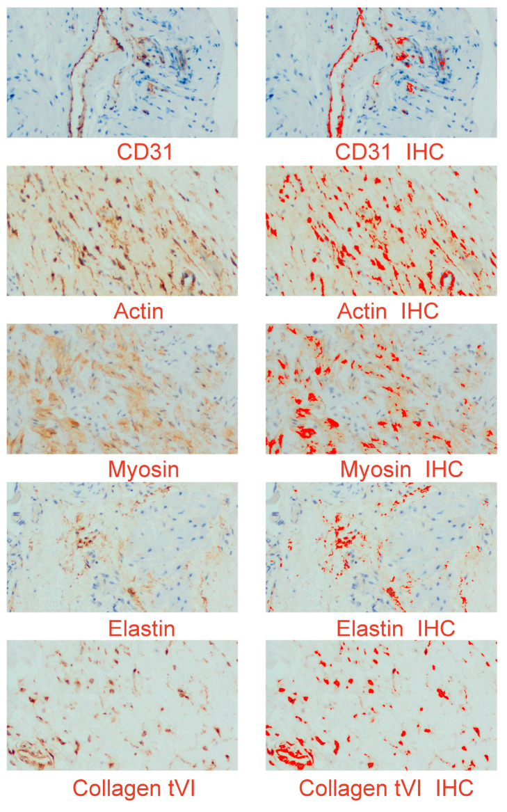
Background and Objectives: Wound healing encompasses a multitude of factors and entails the establishment of interactions among components of the basement membrane. The quantification of particle concentrations can serve as valuable biomarkers for assessing biomechanical muscle properties. The objective of this study was to examine the immunoexpression and immunoconcentration of myometrial collagen type VI, elastin, alpha-smooth muscle actin, and smooth muscle myosin heavy chain, as well as the expression of platelets and clusters of differentiation 31 in the uterine scar following a cesarean section (CS). Materials and Methods: A total of 177 biopsies were procured from a cohort of pregnant women who were healthy, specifically during the surgical procedure of CS. The participants were categorized into seven distinct groups. Group 1 consisted of primiparas, with a total of 52 individuals. The subsequent groups were organized based on the duration of time that had elapsed since their previous CS. The analysis focused on the immunoexpression and immunoconcentration of the particles listed. Results: No significant variations were observed in the myometrial immunoconcentration of collagen type VI, elastin, smooth muscle myosin, and endothelial cell cluster of differentiation 31 among the analyzed groups. The concentration of alpha-smooth muscle actin in the myometrium was found to be significantly higher in patients who underwent CS within a period of less than 2 years since their previous CS, compared to those with a longer interval between procedures. Conclusions: Our findings indicate that the immunoconcentration of uterine myometrial scar collagen type VI, elastin, smooth muscle myosin heavy chain, alpha-smooth muscle actin, and endothelial cell marker cluster of differentiation 31 remains consistent regardless of the duration elapsed since the previous CS. The findings indicate that there are no significant alterations in the biomechanical properties of the uterine muscle beyond a period of 13 months following a CS.
Keywords: cesarean section; cluster of differentiation 31 antigen; immunohistochemistry; uterine cesarean scar.
Conflict of interest statement
The authors declare no conflicts of interest.
Figures
Similar articles
-
Morphological estimation of incomplete uterine scar rupture (dehiscence) in post- cesarean deliveries. Immunohistochemical studies.Ginekol Pol. 2020;91(11):685-692. doi: 10.5603/GP.2020.0115. Ginekol Pol. 2020. PMID: 33301163
-
Uterine wound healing after caesarean section: A systematic review.Eur J Obstet Gynecol Reprod Biol. 2024 May;296:83-90. doi: 10.1016/j.ejogrb.2024.02.045. Epub 2024 Feb 27. Eur J Obstet Gynecol Reprod Biol. 2024. PMID: 38417279
-
Morphology of the cesarean section scar in the non-pregnant uterus after one elective cesarean section.Ginekol Pol. 2017;88(4):174-179. doi: 10.5603/GP.a2017.0034. Ginekol Pol. 2017. PMID: 28509317
-
Does the appearance of the cutaneous scar after cesarean section reflect the residual myometrial thickness?Arch Gynecol Obstet. 2021 Mar;303(3):847-851. doi: 10.1007/s00404-020-05943-2. Epub 2021 Jan 7. Arch Gynecol Obstet. 2021. PMID: 33415438 Free PMC article.
-
Should Cesarean Scar Defect Be Treated Laparoscopically? A Case Report and Review of the Literature.J Minim Invasive Gynecol. 2015 Nov-Dec;22(7):1145-52. doi: 10.1016/j.jmig.2015.06.013. Epub 2015 Jun 26. J Minim Invasive Gynecol. 2015. PMID: 26122897 Review.
References
MeSH terms
Substances
LinkOut - more resources
Full Text Sources
Medical
