Case Reports
. 2024 Apr 19;60(4):655.
doi: 10.3390/medicina60040655.

Noninvasive Ambulatory Electrocardiographic Markers from Patients with COVID-19 Pneumonia: A Report of Three Cases

Affiliations
Case Reports

Noninvasive Ambulatory Electrocardiographic Markers from Patients with COVID-19 Pneumonia: A Report of Three Cases

Motohiro Kimata et al. Medicina (Kaunas). .

Abstract

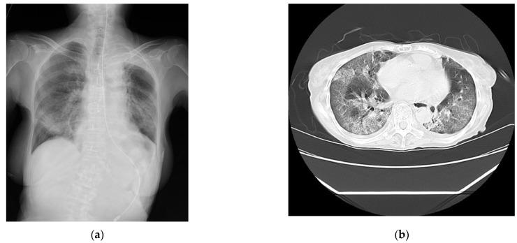
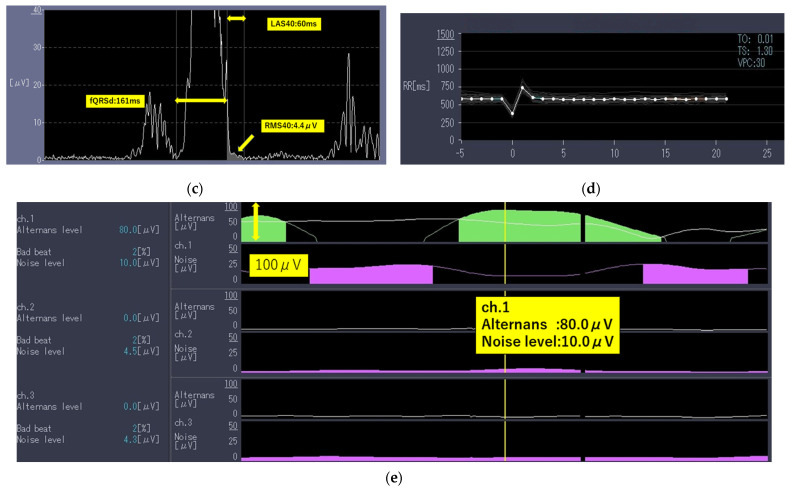
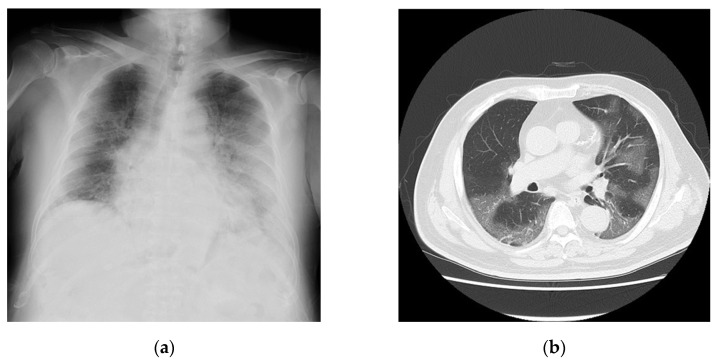
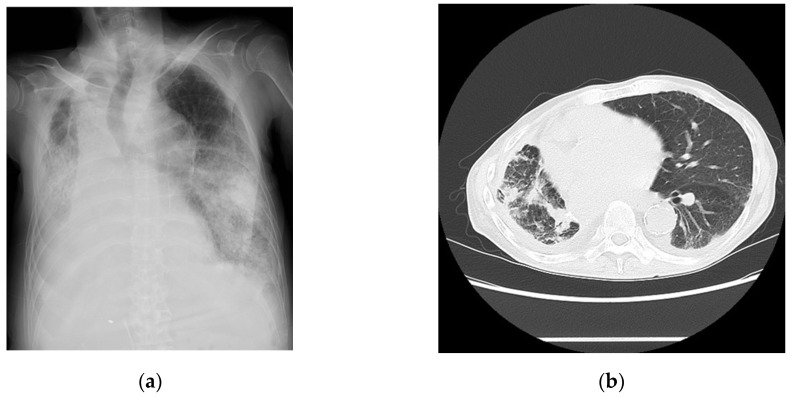
Coronavirus disease 2019 (COVID-19) has affected medical practice. More than 7,000,000 patients died worldwide after being infected with COVID-19; however, no specific laboratory markers have yet been established to predict death related to this disease. In contrast, electrocardiographic changes due to COVID-19 include QT prolongation and ST-T changes; however, there have not been studies on the ambulatory electrocardiographic markers of COVID-19. We encountered three patients diagnosed as having COVID-19 who did not have a prior history of significant structural heart diseases. All patients had abnormalities in ambulatory echocardiogram parameters detected by high-resolution 24 h electrocardiogram monitoring: positive late potentials (LPs) and T-wave alternans (TWA), abnormal heart rate variability (HRV), and heart rate turbulence (HRT). Case 1 involved a 78-year-old woman with a history of chronic kidney disease, Case 2 involved a 76-year-old man with hypertension and diabetes, and Case 3 involved a 67-year-old man with renal cancer, lung cancer, and diabetes. None of them had a prior history of significant structural heart disease. Although no significant consistent increases in clinical markers were observed, all three patients died, mainly because of respiratory failure with mild heart failure. The LP, TWA, HRV, and HRT were positive in all three cases with no significant structural cardiac disease at the initial phase of admission. The further accumulation of data regarding ambulatory electrocardiographic markers in patients with COVID-19 is needed. Depending on the accumulation of data, the LP, TWA, HRV, and HRT could be identified as potential risk factors for COVID-19 pneumonia in the early phase of admission.

Keywords: COVID-19; Holter electrocardiograph; SARS-CoV-2; ambulatory electrocardiographic markers; coronavirus disease-19; global cardiac ischemia; myocardial remodeling; sudden cardiac death.


Conflict of interest statement

The authors declare no conflicts of interest.

Figures

Figure 1
Figure 1
The findings for Case 1. (a) The chest radiograph captured on the day of admission. (b) The chest computed tomography image captured on the day of admission showing bilateral diffuse ground-glass opacities. (c) An analysis of the LPs measured using high-resolution 24 h Holter electrocardiography on the day after admission. The filtered QRS duration (fQRS) was 128 milliseconds, the root-mean-square voltage of the signals (RMS40) was 4.4 μV (<20 μV), and the duration of the low-amplitude signal after the voltage decreased to less than 40 μV (LAS40) was 60 milliseconds (>38 milliseconds). The patient was determined to be positive. (d) An analysis of the heart rate turbulence (HRT) measured using high-resolution 24 h Holter electrocardiography on the day after admission. The turbulence onset was ≥0%, and the turbulence slope was ≤2.5 milliseconds per RR interval. The patient was determined to have abnormal HRT (Category 2). (e) An analysis of T-wave alternans (TWA) measured using high-resolution 24 h Holter electrocardiography on the day after admission. Shaded in green area indicate the presence of TWA. The TWA was 80.0 μV with a noise level of 10.0 mV. The TWA reference value was 19.9 μV when the noise level was less than 10 mV. The patient was determined to be positive.
Figure 1
Figure 1
The findings for Case 1. (a) The chest radiograph captured on the day of admission. (b) The chest computed tomography image captured on the day of admission showing bilateral diffuse ground-glass opacities. (c) An analysis of the LPs measured using high-resolution 24 h Holter electrocardiography on the day after admission. The filtered QRS duration (fQRS) was 128 milliseconds, the root-mean-square voltage of the signals (RMS40) was 4.4 μV (<20 μV), and the duration of the low-amplitude signal after the voltage decreased to less than 40 μV (LAS40) was 60 milliseconds (>38 milliseconds). The patient was determined to be positive. (d) An analysis of the heart rate turbulence (HRT) measured using high-resolution 24 h Holter electrocardiography on the day after admission. The turbulence onset was ≥0%, and the turbulence slope was ≤2.5 milliseconds per RR interval. The patient was determined to have abnormal HRT (Category 2). (e) An analysis of T-wave alternans (TWA) measured using high-resolution 24 h Holter electrocardiography on the day after admission. Shaded in green area indicate the presence of TWA. The TWA was 80.0 μV with a noise level of 10.0 mV. The TWA reference value was 19.9 μV when the noise level was less than 10 mV. The patient was determined to be positive.
Figure 2
Figure 2
Imaging findings of Case 2 captured on day of admission. (a) Chest radiograph and (b) chest computed tomography image showing bilateral diffuse ground-glass opacities.
Figure 3
Figure 3
Imaging findings for Case 3 captured on day of admission. (a) Chest radiograph and (b) chest computed tomography image showing bilateral extensive findings of pneumonia with air bronchogram.

Similar articles

References

    1. World Health Organization Number of COVID-19 deaths reported to WHO. [(accessed on 21 February 2024)]. Available online: https://data.who.int/dashboards/covid19/deaths?n=c.
    1. Sharma Y.P., Agstam S., Yadav A., Gupta A., Gupta A. Cardiovascular manifestations of COVID-19: An evidence-based narrative review. Indian J. Med. Res. 2021;153:7–16. doi: 10.4103/ijmr.IJMR_2450_20. - DOI - PMC - PubMed
    1. Madjid M., Safavi-Naeini P., Solomon S.D., Vardeny O. Potential effects of coronaviruses on the cardiovascular system: A review. JAMA Cardiol. 2020;5:831–840. doi: 10.1001/jamacardio.2020.1286. - DOI - PubMed
    1. Azevedo R.B., Botelho B.G., Hollanda J.V.G., Ferreira L.V.L., Junqueira de Andrade L.Z., Oei S.S.M.L., Mello T.S., Muxfeldt E.S. COVID-19 and the cardiovascular system: A comprehensive review. J. Hum. Hypertens. 2021;35:4–11. doi: 10.1038/s41371-020-0387-4. - DOI - PMC - PubMed
    1. Long B., Brady W.J., Bridwell R.E., Ramzy M., Montrief T., Singh M., Gottlieb M. Electrocardiographic manifestations of COVID-19. Am. J. Emerg. Med. 2021;41:96–103. doi: 10.1016/j.ajem.2020.12.060. - DOI - PMC - PubMed

Publication types