. 2024 Mar 29;12(4):691.
doi: 10.3390/microorganisms12040691.

EnvC Homolog Encoded by Xanthomonas citri subsp. citri Is Necessary for Cell Division and Virulence

Affiliations

EnvC Homolog Encoded by Xanthomonas citri subsp. citri Is Necessary for Cell Division and Virulence

Michelle M Pena et al. Microorganisms. .

Abstract

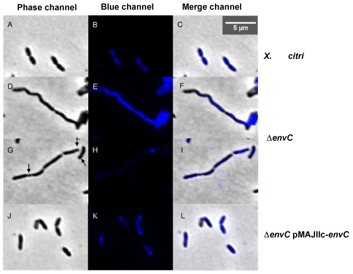
Peptidoglycan hydrolases are enzymes responsible for breaking the peptidoglycan present in the bacterial cell wall, facilitating cell growth, cell division and peptidoglycan turnover. Xanthomonas citri subsp. citri (X. citri), the causal agent of citrus canker, encodes an Escherichia coli M23 peptidase EnvC homolog. EnvC is a LytM factor essential for cleaving the septal peptidoglycan, thereby facilitating the separation of daughter cells. In this study, the investigation focused on EnvC contribution to the virulence and cell separation of X. citri. It was observed that disruption of the X. citri envC gene (ΔenvC) led to a reduction in virulence. Upon inoculation into leaves of Rangpur lime (Citrus limonia Osbeck), the X. citri ΔenvC exhibited a delayed onset of citrus canker symptoms compared with the wild-type X. citri. Mutant complementation restored the wild-type phenotype. Sub-cellular localization confirmed that X. citri EnvC is a periplasmic protein. Moreover, the X. citri ΔenvC mutant exhibited elongated cells, indicating a defect in cell division. These findings support the role of EnvC in the regulation of cell wall organization, cell division, and they clarify the role of this peptidase in X. citri virulence.

Keywords: cell division; citrus canker; peptidoglycan hydrolase; periplasmic protein.


Conflict of interest statement

The authors declare no conflicts of interest.

Figures

Figure 1
Figure 1
Maximum likelihood phylogeny based on the nucleotide sequences of 31 strains representing the Xanthomonas genus. The phylogenetic tree was inferred using RaxMLversion 8.0.24 and drawn using MEGA X software version 10.1.5. Values on the branches indicate bootstrap values for each branch, expressed as percentages. E. coli K12 was used as the outgroup.
Figure 2
Figure 2
Multiple amino acid sequence alignment of M23 peptidase domain for XAC0024 from X. citri 306 (AAM34916.1), XCC0022 from X. campestris (AAM39341.1) and EnvC from E. coli (EGO4467787.1). (A) M23 peptidase domain within the protein sequences of E. coli EnvC, XAC0024 and XCC0022 (purple arrows) and their corresponding locus within genomes (circles on the right). (B) Multiple amino acid sequence alignment of M23 peptidase domain showing matching amino acids for the three organisms. The protein sequences were uploaded from the NCBI Batch Web CD-Search tool. Each protein is depicted with a Peptidase_M13 domain (pfam01551) and a conserved protein domain family EnvC (COG4942). * Conserved sequence (identical); : Conservative mutation; . Semi-conservative mutation.
Figure 3
Figure 3
Pathogenicity assay and growth curve for X. citri 306, ΔenvC and ΔenvC-pMAJIIc-envC. (A) Rangpur lime leaves were infiltrated with cell suspensions of the specified X. citri strains. Pictures were taken at 3, 5, 7, 10, 12 and 15 days after inoculation (DAI). (B) In vitro growth curve. X. citri 306, ΔenvC and ΔenvC-pMAJIIc-envC were cultivated in NB medium, and OD 600 nm readings were taken every 30 min for 72 h. The points on the curves represent the average of triplicate cultures, and the vertical lines indicate the standard deviation values of the means. All experiments were performed in triplicate.
Figure 4
Figure 4
Sub-cellular localization of EnvC-mCherry in X. citri. X. citri strains expressing EnvC-mCherry fusions were cultivated up to OD 600 nm of 0.3, followed by induction with 0.05% arabinose for 2 h before microscope observation. The panels depict the phase contrast images (left), TxRed channels (middle) and the overlay, respectively, for (AC): X. citri pMAJIIc-envC, (DF): X. citri 306. Magnification 100X; scale bar 5 µm.
Figure 5
Figure 5
Cell morphology and nucleoid distribution analyses of X. citri 306, ΔenvC and ΔenvC pMAJIIc-envC strains. The panels show the phase contrast (left), DAPI channels (middle) and the overlay, respectively, for (AC): X. citri 306, (DI): ΔenvC, (HL): ΔenvC-pMAJIIc-envC. Arrows indicate the minicell position. Magnification 100×; scale bar 5 µm.

References

    1. Das A.K. Citrus Canker—A review. J. Appl. Hortic. 2003;5:52–60. doi: 10.37855/jah.2003.v05i01.15. - DOI
    1. Vollmer W., Blanot D., de Pedro M.A. Peptidoglycan structure and architecture. FEMS Microbiol. Rev. 2008;32:149–167. doi: 10.1111/j.1574-6976.2007.00094.x. - DOI - PubMed
    1. Silhavy T.J., Kahne D., Walker S. The bacterial cell envelope. Cold Spring Harb. Perspect. Biol. 2010;2:a000414. doi: 10.1101/cshperspect.a000414. - DOI - PMC - PubMed
    1. Frirdich E., Gaynor E.C. Peptidoglycan hydrolases, bacterial shape, and pathogenesis. Curr. Opin. Microbiol. 2013;16:767–778. doi: 10.1016/j.mib.2013.09.005. - DOI - PubMed
    1. Firczuk M., Bochtler M. Folds and activities of peptidoglycan amidases. FEMS Microbiol. Rev. 2007;31:676–691. doi: 10.1111/j.1574-6976.2007.00084.x. - DOI - PubMed

LinkOut - more resources