. 2024 Apr 18;12(4):820.
doi: 10.3390/microorganisms12040820.

Ligand-Free Silver Nanoparticles: An Innovative Strategy against Viruses and Bacteria

Affiliations

Ligand-Free Silver Nanoparticles: An Innovative Strategy against Viruses and Bacteria

Maria Vittoria Morone et al. Microorganisms. .

Abstract

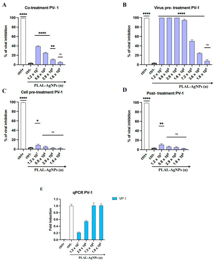
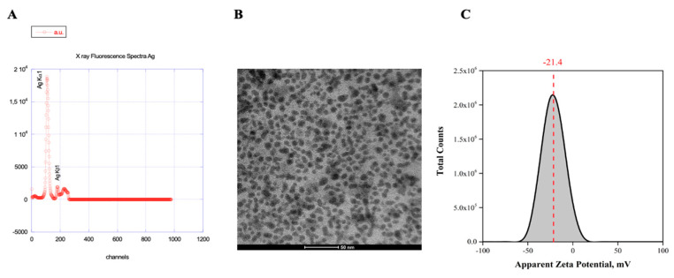
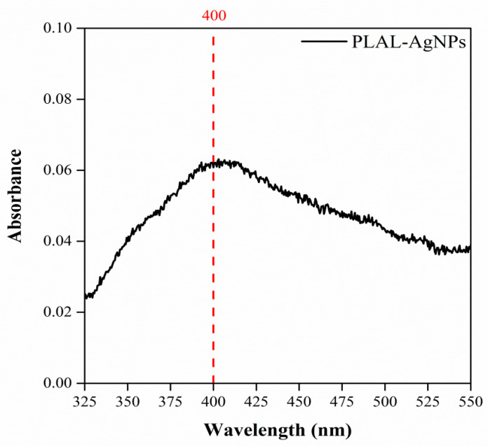
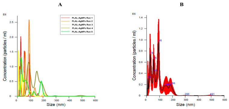
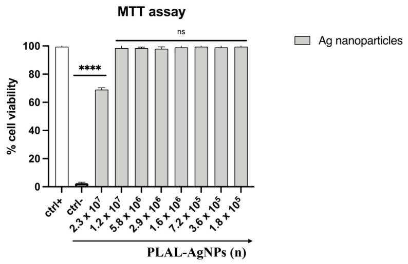
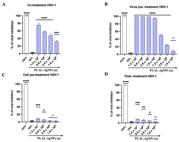
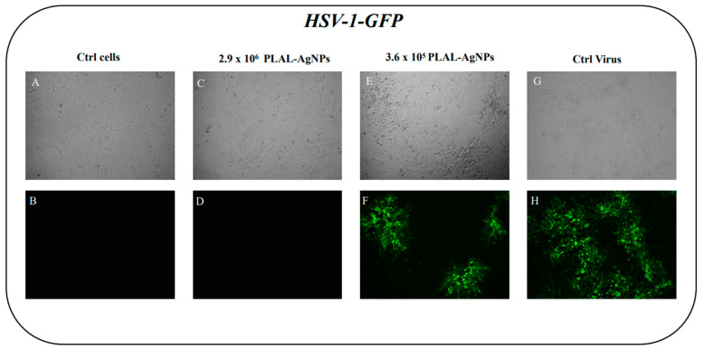
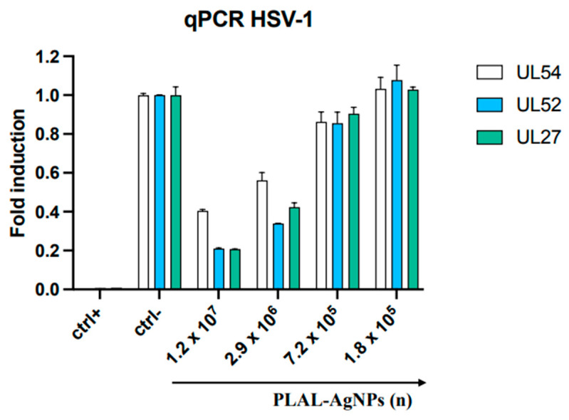
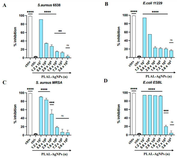
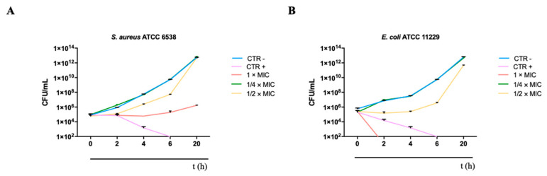
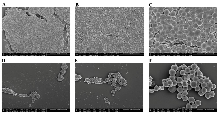
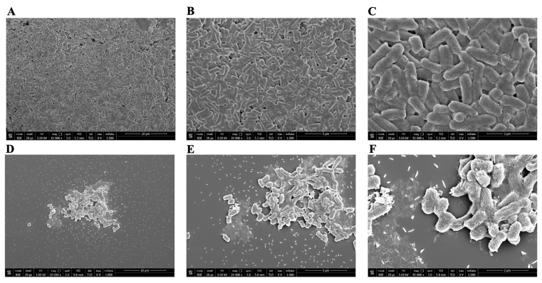
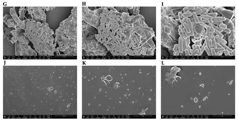
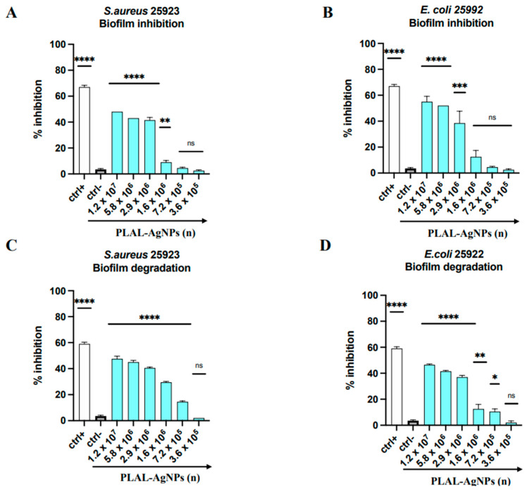
The spread of antibiotic-resistant bacteria and the rise of emerging and re-emerging viruses in recent years constitute significant public health problems. Therefore, it is necessary to develop new antimicrobial strategies to overcome these challenges. Herein, we describe an innovative method to synthesize ligand-free silver nanoparticles by Pulsed Laser Ablation in Liquid (PLAL-AgNPs). Thus produced, nanoparticles were characterized by total X-ray fluorescence, zeta potential analysis, transmission electron microscopy (TEM), and nanoparticle tracking analysis (NTA). A 3-(4,5-dimethylthiazol-2-yl)-2,5-diphenyltetrazolium bromide (MTT) assay was performed to evaluate the nanoparticles' cytotoxicity. Their potential was evaluated against the enveloped herpes simplex virus type 1 (HSV-1) and the naked poliovirus type 1 (PV-1) by plaque reduction assays and confirmed by real-time PCR and fluorescence microscopy, showing that nanoparticles interfered with the early stage of infection. Their action was also examined against different bacteria. We observed that the PLAL-AgNPs exerted a strong effect against both methicillin-resistant Staphylococcus aureus (S. aureus MRSA) and Escherichia coli (E. coli) producing extended-spectrum β-lactamase (ESBL). In detail, the PLAL-AgNPs exhibited a bacteriostatic action against S. aureus and a bactericidal activity against E. coli. Finally, we proved that the PLAL-AgNPs were able to inhibit/degrade the biofilm of S. aureus and E. coli.

Keywords: PLAL-AgNPs; antibacterial activity; antibiofilm activity; antiviral activity; herpesvirus; multidrug resistance; poliovirus; silver nanoparticles.


Conflict of interest statement

The authors declare no conflicts of interest.

Figures

Figure 8
Figure 8
Antiviral activity of PLAL-AgNPs against PV-1 infection: (A) co-treatment; (B) virus pre-treatment; (C) cell pre-treatment; (D) post-treatment. Different compounds were used as positive controls (ctr+) for each treatment: pleconaril ((A,B), 2 μg/mL for both assays) [37], WIN51711 ((C) 5 μg/mL), and protein 2C ((D), 10 μM) [38], while infected cells were used as negative controls (ctr-). Data represent the means ± standard deviations (SDs) of three independent experiments. **** < 0.0001, ** = 0.0032, * = 0.0457; ns = non-significant. (E) Molecular assay. Real-time PCR was performed to evaluate the effect of AgNPs on viral gene expression. Virus pre-treatment assay was performed, RNA was extracted after 24 h, then retrotranscribed into cDNA and amplified. Gene expression of the capsid protein VP1 was evaluated. Infected cells represent the ctrl-, while uninfected cells represent the ctrl+.
Figure 1
Figure 1
Physical–chemical characterization. (A) X-ray fluorescence spectrum of Ag. The characteristic X-ray emission lines of the Ag target used to produce PLAL-AgNPs are present, while no other material is visible, demonstrating the purity of the Ag target. (B) TEM visualization of PLAL-AgNPs at 50 nm magnification. (C) Zeta potential analysis of PLAL-AgNPs. The zeta potential value of the PLAL-AgNPs is −21.4 mV.
Figure 2
Figure 2
UV-vis absorption spectrum of PLAL-AgNPs in phosphate-buffered solution at 25 °C. PLAL-AgNPs showed a surface plasmon resonance band at approximately 400 nm.
Figure 3
Figure 3
Nanoparticle tracking analysis (NTA) of PLAL-AgNPs in a 1 mL sample. (A) Hydrodynamic diameter size per concentration, obtained using the finite track length adjustment (FTLA) algorithm, with quintuplicate measurements (one to five runs) of the PLAL-AgNPs sample. (B) Average of the five measurements. All measurements were acquired using a dilution of 1:4 in PBS 1×. All the data indicate good sample reproducibility. Black line: medium value of the five measurements; red line: standard deviation.
Figure 4
Figure 4
Cytotoxicity of PLAL-AgNPs on Vero cells. The percentage of cell viability was evaluated by MTT assay. The cell monolayers were treated with NPs (from 2.3 × 10⁷ to 1.8 × 10⁵) for 24 h. Untreated cells correspond to the ctrl+ of the experiment, whereas DMSO (100%) was used as a negative control (ctrl-). Data represent means ± standard deviations (SDs) of three independent experiments. ****: p-value < 0.0001; ns: non-significant.
Figure 5
Figure 5
Antiviral activity of PLAL-AgNPs against HSV-1 infection: (A) co-treatment; (B) virus pre-treatment; (C) cell pre-treatment; (D) post-treatment. Different compounds were used as a positive control (ctr+) for each treatment: melittin ((A,B), 5 μM for both assays), dextran sulfate ((C), 1 μM), and aciclovir ((D), 5 μM) [34]. At the same time, the infected cells were used as a negative control (ctrl-). Data represent the means ± standard deviations (SDs) of three independent experiments. **** < 0.0001, *** = 0.0002, ** = 0.0015, * = 0.0191; ns: non-significant.
Figure 6
Figure 6
Antiviral activity of PLAL-AgNPs against HSV-1-GFP. Fluorescent and RGB images are shown. (A,B) Untreated and uninfected cells. (C,D) No plaques or GFP signals were present when cells were treated with 2.9 × 10⁶ PLAL-AgNPs. (E,F) Plaques in cells treated with 3.6 × 10⁵ AgNPs. (G,H) Control infected cells.
Figure 7
Figure 7
Molecular assay. Real-time PCR was conducted to evaluate the effect of AgNPs on viral gene expression. Virus pre-treatment assay was performed, RNA was extracted after 30 h, then it was retrotranscribed into cDNA and amplified. The expression of UL54, UL52, and UL27 was analyzed. Infected cells represent the ctrl-; uninfected cells represent the ctrl+.
Figure 9
Figure 9
Antibacterial activity of PLAL-AgNPs against (A) S. aureus 6538, (B) E. coli 11229, (C) S. aureus MDR, and (D) E. coli ESBL. Statistical significance was referred to the negative control and determined by analysis of variance (ANOVA). Dunnett’s test was used for multiple comparisons with controls. ****: p-value < 0.0001, ***: p-value = 0.0004, **: p-value = 0.0055; ns: p-value = non-significant.
Figure 10
Figure 10
Killing kinetics of AgNPs against (A) S. aureus 6538 and (B) E. coli 11229.
Figure 11
Figure 11
SEM visualization of PLAL-AgNPs and S. aureus. (AC) Untreated S. aureus 6538 cells (ctrl-) at a magnification of 10,000, 20,000, and 50,000×, respectively. (DF) S. aureus treated with 1.2 × 10⁷ PLAL-AgNPs (MIC) at a magnification of 10,000, 20,000, and 50,000×, respectively. (GI) S. aureus treated with 5.8 × 10⁶ PLAL-AgNPs (½ MIC) at a magnification of 10,000, 20,000, and 50,000×, respectively. (JL) S. aureus treated with 2 μg/mL of vancomycin (ctrl+) at a magnification of 10,000, 20,000, and 50,000×, respectively.
Figure 11
Figure 11
SEM visualization of PLAL-AgNPs and S. aureus. (AC) Untreated S. aureus 6538 cells (ctrl-) at a magnification of 10,000, 20,000, and 50,000×, respectively. (DF) S. aureus treated with 1.2 × 10⁷ PLAL-AgNPs (MIC) at a magnification of 10,000, 20,000, and 50,000×, respectively. (GI) S. aureus treated with 5.8 × 10⁶ PLAL-AgNPs (½ MIC) at a magnification of 10,000, 20,000, and 50,000×, respectively. (JL) S. aureus treated with 2 μg/mL of vancomycin (ctrl+) at a magnification of 10,000, 20,000, and 50,000×, respectively.
Figure 12
Figure 12
SEM visualization of PLAL-AgNPs and E. coli. (AC) Untreated E. coli 11229 cells (ctrl-) at a magnification of 10,000, 20,000, and 50,000×, respectively. (DF) E. coli treated with 1.2 × 10⁷ PLAL-AgNPs (MIC) at a magnification of 10,000, 20,000, and 50,000×, respectively. (GI) E. coli treated with 5.8 × 10⁶ PLAL-AgNPs (½ MIC) at a magnification of 10,000, 20,000, and 50,000×, respectively. (JL) E. coli cells treated with 8 μg/mL of amikacin (ctrl+) at a magnification of 10,000, 20,000, and 50,000×, respectively.
Figure 12
Figure 12
SEM visualization of PLAL-AgNPs and E. coli. (AC) Untreated E. coli 11229 cells (ctrl-) at a magnification of 10,000, 20,000, and 50,000×, respectively. (DF) E. coli treated with 1.2 × 10⁷ PLAL-AgNPs (MIC) at a magnification of 10,000, 20,000, and 50,000×, respectively. (GI) E. coli treated with 5.8 × 10⁶ PLAL-AgNPs (½ MIC) at a magnification of 10,000, 20,000, and 50,000×, respectively. (JL) E. coli cells treated with 8 μg/mL of amikacin (ctrl+) at a magnification of 10,000, 20,000, and 50,000×, respectively.
Figure 13
Figure 13
Antibiofilm activity of PLAL-AgNPs. Biofilm inhibition and degradation against S. aureus (A,C). Biofilm inhibition and degradation against E. coli (B,D). Data represent the means ± standard deviations (SDs) of three independent experiments. ****: p-value < 0.0001, ***: p-value = 0.0001, **: p-value = 0.0058, *: p-value = 0.0159; ns: non-significant.

Similar articles

Cited by

References

    1. Van Seventer J.M., Hochberg N.S. International Encyclopedia of Public Health. Elsevier; Amsterdam, The Netherlands: 2017. Principles of Infectious Diseases: Transmission, Diagnosis, Prevention, and Control; pp. 22–39.
    1. Ismahene Y. Infectious Diseases, Trade, and Economic Growth: A Panel Analysis of Developed and Developing Countries. J. Knowl. Econ. 2022;13:2547–2583. doi: 10.1007/s13132-021-00811-z. - DOI
    1. Salam M.A., Al-Amin M.Y., Salam M.T., Pawar J.S., Akhter N., Rabaan A.A., Alqumber M.A.A. Antimicrobial Resistance: A Growing Serious Threat for Global Public Health. Healthcare. 2023;11:1946. doi: 10.3390/healthcare11131946. - DOI - PMC - PubMed
    1. Jiang Y.-C., Feng H., Lin Y.-C., Guo X.-R. New strategies against drug resistance to herpes simplex virus. Int. J. Oral Sci. 2016;8:1–6. doi: 10.1038/ijos.2016.3. - DOI - PMC - PubMed
    1. Anton-Vazquez V., Mehra V., Mbisa J.L., Bradshaw D., Basu T.N., Daly M.-L., Mufti G.J., Pagliuca A., Potter V., Zuckerman M. Challenges of aciclovir-resistant HSV infection in allogeneic bone marrow transplant recipients. J. Clin. Virol. 2020;128:104421. doi: 10.1016/j.jcv.2020.104421. - DOI - PubMed

LinkOut - more resources