

Association of Acidotolerant Cyanobacteria to Microbial Mats below pH 1 in Acidic Mineral Precipitates in Río Tinto River in Spain
- PMID: 38674771
- PMCID: PMC11052175
- DOI: 10.3390/microorganisms12040829
Association of Acidotolerant Cyanobacteria to Microbial Mats below pH 1 in Acidic Mineral Precipitates in Río Tinto River in Spain
Abstract
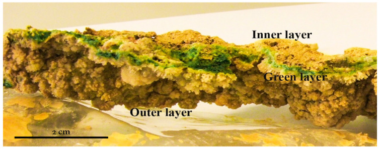
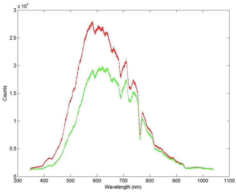
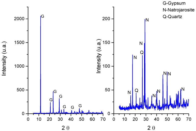
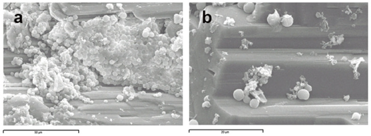
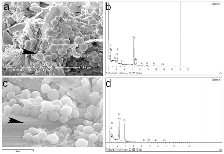
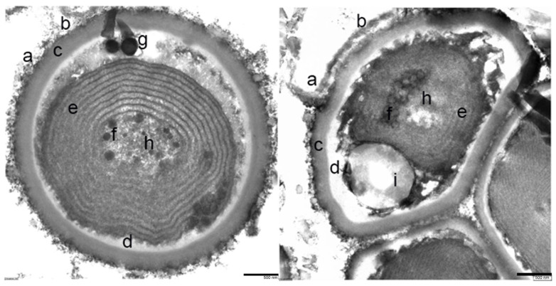
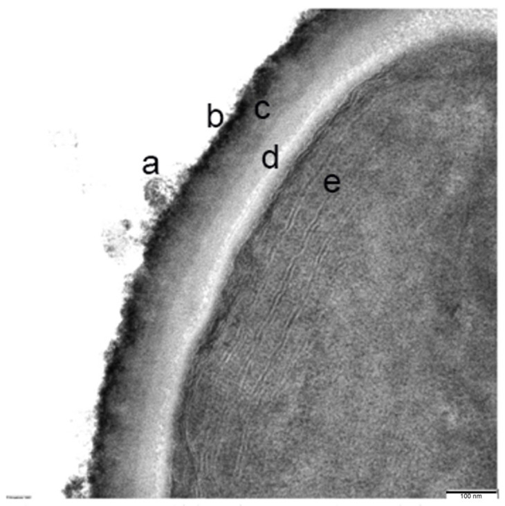
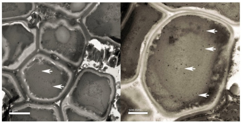
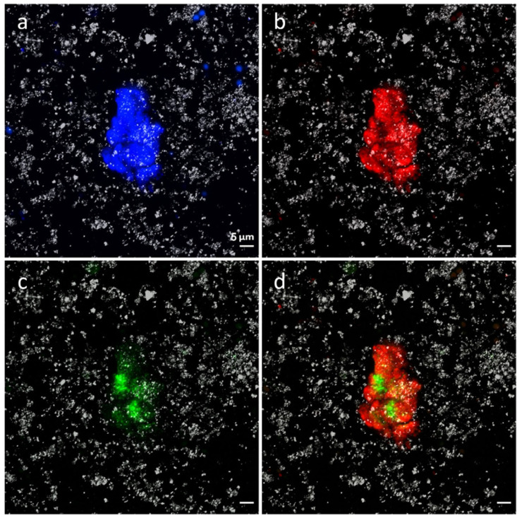
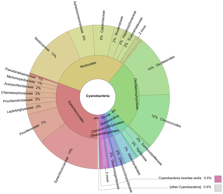
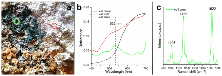
This report describes acidic microbial mats containing cyanobacteria that are strongly associated to precipitated minerals in the source area of Río Tinto. Río Tinto (Huelva, Southwestern Spain) is an extreme acidic environment where iron and sulfur cycles play a fundamental role in sustaining the extremely low pH and the high concentration of heavy metals, while maintaining a high level of microbial diversity. These multi-layered mineral deposits are stable all year round and are characterized by a succession of thick greenish-blue and brownish layers mainly composed of natrojarosite. The temperature and absorbance above and below the mineral precipitates were followed and stable conditions were detected inside the mineral precipitates. Different methodologies, scanning and transmission electron microscopy, immunological detection, fluorescence in situ hybridization, and metagenomic analysis were used to describe the biodiversity existing in these microbial mats, demonstrating, for the first time, the existence of acid-tolerant cyanobacteria in a hyperacidic environment of below pH 1. Up to 0.46% of the classified sequences belong to cyanobacterial microorganisms, and 1.47% of the aligned DNA reads belong to the Cyanobacteria clade.
Keywords: acidophiles; astrobiology; cyanobacteria; earth analogues; endolithic ecosystems; extremophiles; natrojarosite.
Conflict of interest statement
The authors declare no conflicts of interest. The funders had no role in the design of the study; in the collection, analyses, or interpretation of data; in the writing of the manuscript; or in the decision to publish the results.
Figures
Similar articles
-
Biogeochemical Niches of Fe-Cycling Communities Influencing Heavy Metal Transport along the Rio Tinto, Spain.Appl Environ Microbiol. 2022 Feb 22;88(4):e0229021. doi: 10.1128/AEM.02290-21. Epub 2021 Dec 15. Appl Environ Microbiol. 2022. PMID: 34910570 Free PMC article.
-
Río tinto: a geochemical and mineralogical terrestrial analogue of Mars.Life (Basel). 2014 Sep 15;4(3):511-34. doi: 10.3390/life4030511. Life (Basel). 2014. PMID: 25370383 Free PMC article. Review.
-
Eukaryotic organisms in extreme acidic environments, the río tinto case.Life (Basel). 2013 Jul 4;3(3):363-74. doi: 10.3390/life3030363. Life (Basel). 2013. PMID: 25369810 Free PMC article. Review.
-
Microbial diversity in anaerobic sediments at Rio Tinto, a naturally acidic environment with a high heavy metal content.Appl Environ Microbiol. 2011 Sep;77(17):6085-93. doi: 10.1128/AEM.00654-11. Epub 2011 Jul 1. Appl Environ Microbiol. 2011. PMID: 21724883 Free PMC article.
-
Comparative microbial ecology of the water column of an extreme acidic pit lake, Nuestra Señora del Carmen, and the Río Tinto basin (Iberian Pyrite Belt).Int Microbiol. 2014 Dec;17(4):225-33. doi: 10.2436/20.1501.01.225. Int Microbiol. 2014. PMID: 26421738
References
-
- Whitton B.A., Potts M., editors. Ecology of Cyanobacteria II: Their Diversity in Space and Time. Springer Science & Business Media; Berlin/Heidelberg, Germany: 2012.
LinkOut - more resources
Full Text Sources
