. 2024 Apr 9;16(8):1100.
doi: 10.3390/nu16081100.

Faecalibacterium prausnitzii Supplementation Prevents Intestinal Barrier Injury and Gut Microflora Dysbiosis Induced by Sleep Deprivation

Affiliations

Faecalibacterium prausnitzii Supplementation Prevents Intestinal Barrier Injury and Gut Microflora Dysbiosis Induced by Sleep Deprivation

Xintong Wang et al. Nutrients. .

Abstract

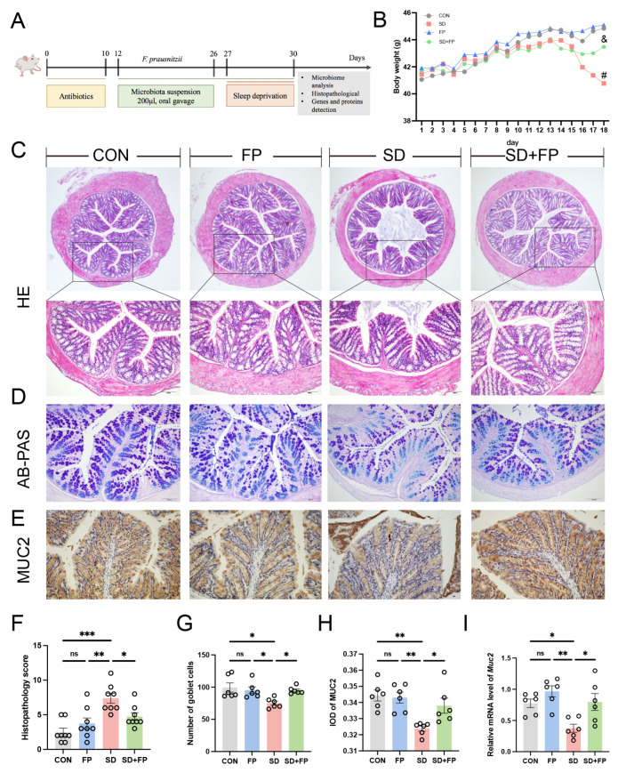
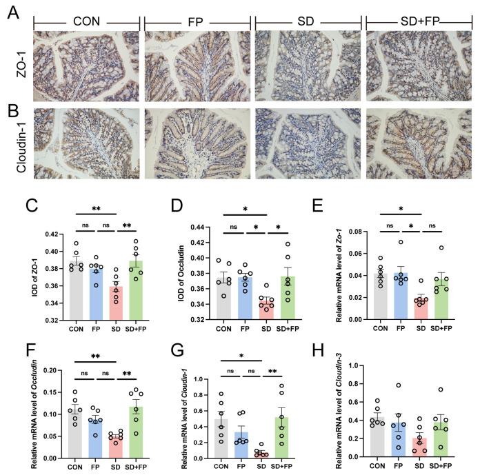
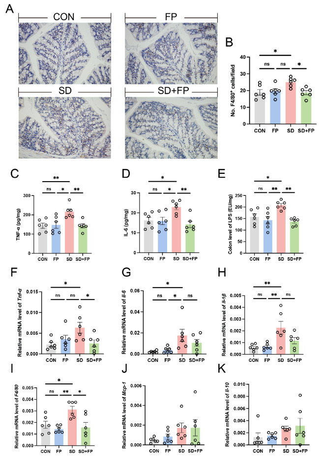
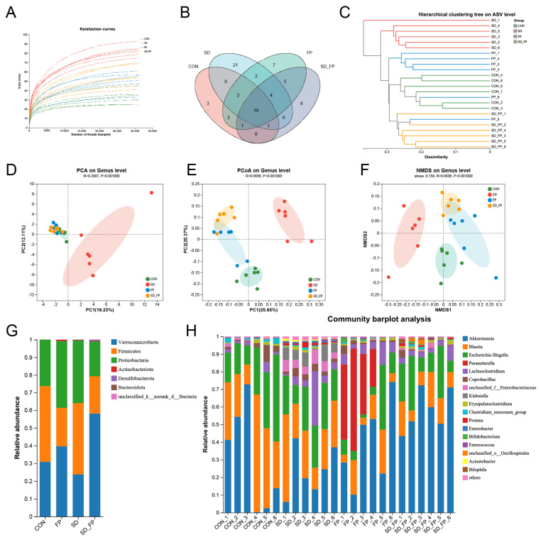
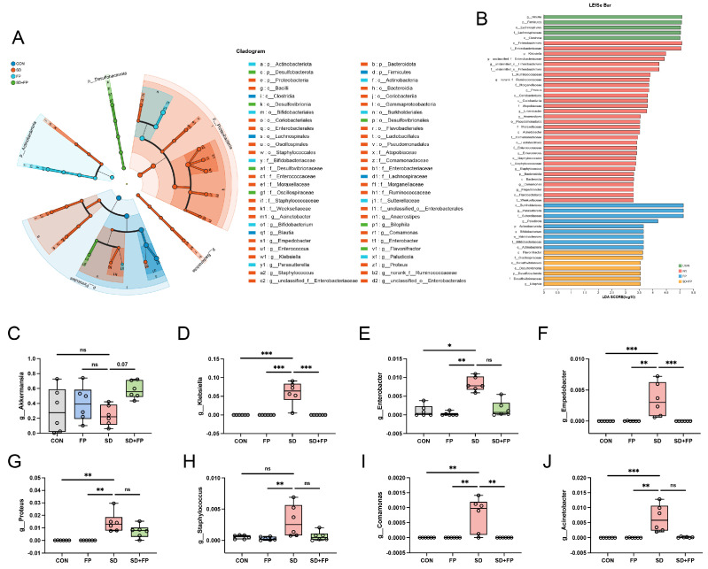
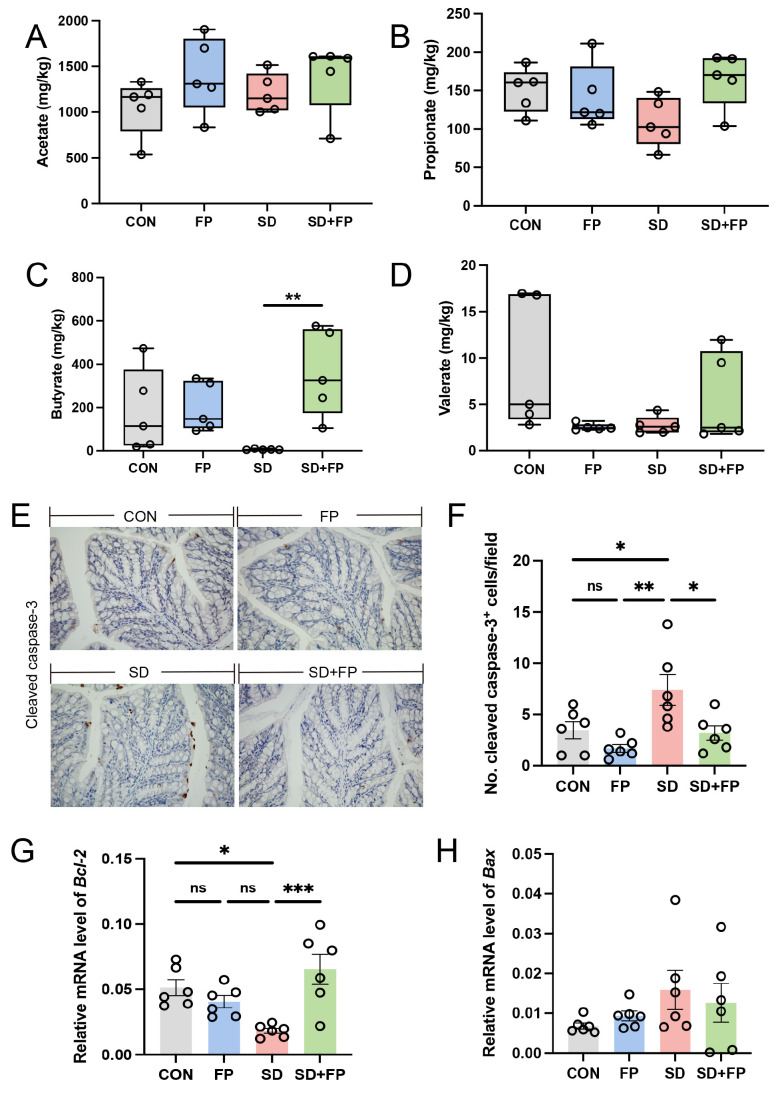
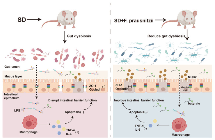
Sleep deprivation (SD) leads to impaired intestinal barrier function and intestinal flora disorder, especially a reduction in the abundance of the next generation of probiotic Faecalibacterium prausnitzii (F. prausnitzii). However, it remains largely unclear whether F. prausnitzii can ameliorate SD-induced intestinal barrier damage. A 72 h SD mouse model was used in this research, with or without the addition of F. prausnitzii. The findings indicated that pre-colonization with F. prausnitzii could protect against tissue damage from SD, enhance goblet cell count and MUC2 levels in the colon, boost tight-junction protein expression, decrease macrophage infiltration, suppress pro-inflammatory cytokine expression, and reduce apoptosis. We found that the presence of F. prausnitzii helped to balance the gut microbiota in SD mice by reducing harmful bacteria like Klebsiella and Staphylococcus, while increasing beneficial bacteria such as Akkermansia. Ion chromatography analysis revealed that F. prausnitzii pretreatment increased the fecal butyrate level in SD mice. Overall, these results suggested that incorporating F. prausnitzii could help reduce gut damage caused by SD, potentially by enhancing the intestinal barrier and balancing gut microflora. This provides a foundation for utilizing probiotics to protect against intestinal illnesses.

Keywords: Faecalibacterium prausnitzii; intestinal barrier function; intestinal microflora; short chain fatty acids; sleep deprivation.


Conflict of interest statement

The authors declare no conflicts of interest.

Figures

Figure 1
Figure 1
Faecalibacterium prausnitzii colonization alleviated intestinal mucosal barrier disruption induced by SD. (A) Diagram showing the layout of the experiment. (B) Body weight. (C) Hematoxylin and eosin (H&E) staining, Bar = 100 μm. (D) Alcian blue, and periodic acid-Schiff (AB-PAS) staining, Bar = 100 μm. (E) Representative captures of immunohistochemical of MUC2 in colon, Bar = 100 μm. (F) Histologic scores (n = 8). (G) The number of goblet cells per crypt (n = 6). (H) IOD of MUC2 in the intestinal tissue from each treatment group (n = 6). (I) The mRNA levels of Muc2 in the colon (n = 6). One-way ANOVA was utilized to evaluate variances. The study included a control group (CON), a group colonized with Faecalibacterium prausnitzii (FP), a group subjected to sleep deprivation (SD), and a group experiencing sleep deprivation with Faecalibacterium prausnitzii colonization (SD + FP). The outcome indicates the average value plus or minus the standard error. *, p < 0.05; **, p < 0.01; ***, p < 0.001 compared with the control group. ns, non-significance. # p < 0.05 vs. the CON group; & p < 0.05 vs. the SD group.
Figure 2
Figure 2
Faecalibacterium prausnitzii colonization increased the expression of tight-junction proteins in sleep-deprived mice. (A) Representative captures of immunohistochemical of ZO-1 in colon, Bar = 100 μm. (B) Representative captures of immunohistochemical of occludin in colon, Bar = 100 μm. (C) IOD of ZO-1 in the intestinal tissue from each treatment group (n = 6). (D) IOD of occludin in the intestinal tissue from each treatment group (n = 6). (EH) The mRNA levels of ZO-1, occludin, Cloudin-1, and Cloudin-3 in the colon (n = 6). One-way ANOVA was utilized to evaluate variances. The study included a control group (CON), a group colonized with Faecalibacterium prausnitzii (FP), a group subjected to sleep deprivation (SD), and a group experiencing sleep deprivation with Faecalibacterium prausnitzii colonization (SD + FP). The outcome indicates the average value plus or minus the standard error. *, p < 0.05; **, p < 0.01 compared with the control group. ns, non-significance.
Figure 3
Figure 3
Faecalibacterium prausnitzii colonization inhibited the expression of inflammatory cytokines in sleep-deprived mice. (A) Representative captures of immunohistochemical of F4/80 in colon, Bar = 100 μm. (B) The quantification of F4/80+ cells (n = 6). (C,D) The levels of cytokines (TNF-α and IL-6) in the colon (n = 6). (E) The levels of LPS in the colon (n = 6). (FK) The mRNA levels of Tnf-α, Il-6, Il-1β, F4/80, Mcp-1 and Il-10 in the colon (n = 6). One-way ANOVA was utilized to evaluate variances. The study included a control group (CON), a group colonized with Faecalibacterium prausnitzii (FP), a group subjected to sleep deprivation (SD), and a group experiencing sleep deprivation with Faecalibacterium prausnitzii colonization (SD + FP). The outcome indicates the average value plus or minus the standard error. *, p < 0.05; **, p < 0.01 compared with the control group. ns, non-significance.
Figure 4
Figure 4
Composition of the colonic microbiota in mice. (A) Rarefaction curves. (B) Venn diagram. (C) Unweighted pair-group method with arithmetic mean (UPGMA) analysis (at the genus level). (D) Principal component analysis (PCA). (E) PCoA score plot. (F) Nonmetric multidimensional scaling (NMDS) score plot based on the binary_jaccard distance plot based on the ASV of the gut microbe. (G) Relative abundances of gut microbiota at the phylum level. (H) Relative abundances of gut microbiota at the genus level. The study included a control group (CON), a group colonized with Faecalibacterium prausnitzii (FP), a group subjected to sleep deprivation (SD), and a group experiencing sleep deprivation with Faecalibacterium prausnitzii colonization (SD + FP).
Figure 5
Figure 5
Faecalibacterium prausnitzii colonization inhibited the colonic microbial dysbiosis induced by sleep deprivation. (A) Taxonomic cladogram obtained from LEfSe sequence analysis in the colon. Biomarker taxa are highlighted by colored circles and shaded areas. (B) The diameter of each circle reflects the abundance of that taxon in the community. A cutoff value of 3 was used for LDA. (CJ) Relative abundance of g__Akkermansia, g__Klebsiella, g__Enterobacter, g__Enterobacter, g__Proteus, g__Staphylococcus, g__Comamonas, g__Acinetobacter in the colon microbiota based on the LefSe results. The study included a control group (CON), a group colonized with Faecalibacterium prausnitzii (FP), a group subjected to sleep deprivation (SD), and a group experiencing sleep deprivation with Faecalibacterium prausnitzii colonization (SD + FP). The outcome indicates the average value plus or minus the standard error. *, p < 0.05; **, p < 0.01; ***, p < 0.001 compared with the control group. ns, non-significance.
Figure 6
Figure 6
Faecalibacterium prausnitzii colonization reversed decrease in SCFAs and increase in intestinal apoptosis induced by sleep deprivation. (A) The colon acetate concentration (n = 5). (B) The colon propionate concentration (n = 5). (C) The colon butyrate concentration (n = 5). (D) The colon valerate concentration (n = 5). (E) Representative captures of immunohistochemical of cleaved caspase-3 in colon, Bar = 100 μm. (F) The quantification of cleaved caspase-3+ cells (n = 6). (G,H) The mRNA levels of Bax and Bcl-2 in the colon (n = 6). One-way ANOVA was utilized to evaluate variances. The study included a control group (CON), a group colonized with Faecalibacterium prausnitzii (FP), a group subjected to sleep deprivation (SD), and a group experiencing sleep deprivation with Faecalibacterium prausnitzii colonization (SD + FP). The outcome indicates the average value plus or minus the standard error. *, p < 0.05; **, p < 0.01; ***, p < 0.001 compared with the control group. ns, non-significance.
Figure 7
Figure 7
Schematic diagram of the potential mechanism by which Faecalibacterium prausnitzii ameliorates intestinal injury induced by sleep deprivation. A feasible mechanism is that the colonization of Faecalibacterium prausnitzii ameliorates impaired intestinal barrier function in SD mice through regulating intestinal inflammation, programmed death, and the gut microbiota. SD: sleep deprivation; LPS: lipopolysaccharide; ZO-1: tight-junction protein zonula occluden-1; TNF-α: tumor necrosis factor-alpha; IL-6; interleukin-6. (+); promote; (-); inhibit.

Similar articles

Cited by

References

    1. Kroese F.M., Evers C., Adriaanse M.A., de Ridder D.T.D. Bedtime procrastination: A self-regulation perspective on sleep insufficiency in the general population. J. Health Psychol. 2016;21:853–862. doi: 10.1177/1359105314540014. - DOI - PubMed
    1. Zamore Z., Veasey S.C. Neural consequences of chronic sleep disruption. Trends Neurosci. 2022;45:678–691. doi: 10.1016/j.tins.2022.05.007. - DOI - PMC - PubMed
    1. Neroni B., Evangelisti M., Radocchia G., Di Nardo G., Pantanella F., Villa M.P., Schippa S. Relationship between sleep disorders and gut dysbiosis: What affects what? Sleep. Med. 2021;87:1–7. doi: 10.1016/j.sleep.2021.08.003. - DOI - PubMed
    1. Zeng Y., Zhang Z., Liang S., Chang X., Qin R., Chen H., Guo L. Paternal sleep deprivation induces metabolic perturbations in male offspring via altered LRP5 DNA methylation of pancreatic islets. J. Pineal Res. 2023;74:e12863. doi: 10.1111/jpi.12863. - DOI - PubMed
    1. Fang D., Xu T., Sun J., Shi J., Li F., Yin Y., Wang Z., Liu Y. Nicotinamide Mononucleotide Ameliorates Sleep Deprivation-Induced Gut Microbiota Dysbiosis and Restores Colonization Resistance against Intestinal Infections. Adv. Sci. 2023;10:e2207170. doi: 10.1002/advs.202207170. - DOI - PMC - PubMed

LinkOut - more resources