. 2024 Apr 19;16(8):1212.
doi: 10.3390/nu16081212.

Novel Fermentates Can Enhance Key Immune Responses Associated with Viral Immunity

Affiliations

Novel Fermentates Can Enhance Key Immune Responses Associated with Viral Immunity

Dearbhla Finnegan et al. Nutrients. .

Abstract

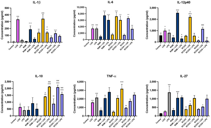
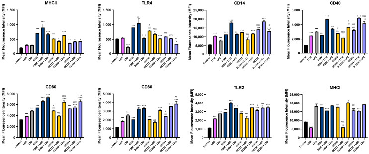
Fermented foods have long been known to have immunomodulatory capabilities, and fermentates derived from the lactic acid bacteria of dairy products can modulate the immune system. We have used skimmed milk powder to generate novel fermentates using Lb. helveticus strains SC234 and SC232 and we demonstrate here that these fermentates can enhance key immune mechanisms that are critical to the immune response to viruses. We show that our novel fermentates, SC234 and SC232, can positively impact on cytokine and chemokine secretion, nitric oxide (NO) production, cell surface marker expression, and phagocytosis in macrophage models. We demonstrate that the fermentates SC234 and SC232 increase the secretion of cytokines IL-1β, IL-6, TNF-α, IL-27, and IL-10; promote an M1 pro-inflammatory phenotype for viral immunity via NO induction; decrease chemokine expression of Monocyte Chemoattractant Protein (MCP); increase cell surface marker expression; and enhance phagocytosis in comparison to their starting material. These data suggest that these novel fermentates have potential as novel functional food ingredients for the treatment, management, and control of viral infection.

Keywords: fermentates; functional food; immune boosting; immunomodulation; macrophage; viral immunity.


Conflict of interest statement

Authors Monica A. Mechoud, Tom Beresford, Harsh Mathur, Paul D. Cotter were employed by the company Teagasc. The remaining authors declare that the research was conducted in the absence of any commercial or financial relationships that could be construed as a potential conflict of interest.

Figures

Figure 1
Figure 1
Exposure of LOX- and LPS-activated BMDM to 25 mg/mL fermentates results in the secretion of cytokines. BMDM cells were seeded at 1 × 10⁶ cells/mL and incubated overnight at 37 °C in 5% CO2. The following day, cells were stimulated with 25 mg/mL fermentate, incubated for 1 h at 37 °C in 5% CO2 and subsequently exposed to LOX 0.5 mM; LPS 100 ng/mL before incubating overnight under the same conditions. Non-fermented RSM was the fermentate control. Supernatants were removed after 24 h and ELISA was performed for cytokines IL-1β, IL-6, IL-10, TNF-α, IL-12p40, and IL-27. Data are presented as mean ± SEM of three replicates. Significance determined using one-way ANOVA with a Newman–Keuls post-test. Output p value style APA: 0.12 nonsignificant (unlabelled), 0.033 somewhat significant (*), 0.002 significant (**), and <0.001 highly significant (***); where the following symbols represent; (1) comparing control cells to LOX and LPS and unstimulated samples “*”, (2) comparing TLR to sample + TLR “+”, and (3) comparing RSM +/− TLR to sample +/− TLR “x”.
Figure 2
Figure 2
Exposure of M1/M2-polarised BMDMs to 25 mg/mL fermentates results in the secretion of cytokines IL-6, TNF-α, and IL-10. BMDM cells were seeded at 5 × 10⁵ cells/mL and incubated for 1 h at 37 °C in 5% CO2. Cells were stimulated with 25 mg/mL fermentates, incubated for 3 h at 37 °C in 5% CO2. The cells were either polarised to the M1 phenotype by stimulating with LPS (100 ng/mL) in the presence of 20 ng/mL rIFN-γ or towards M2 cells by adding 20 ng/mL rIL-4, 20 ng/mL IL-13, and 20 ng/mL rTGF-B and incubating for 24 h at 37 °C. Supernatants were removed after 24 h and ELISA was performed for IL-6, IL-10, and TNF-α. Data are presented as mean ± SEM of three replicates. (A) represent the M0, M1, and M2 profiles for each cytokine. (BD) represent cytokine output in response to sample presence. Significance determined using one-way ANOVA with a Newman–Keuls post-test. Output p value style APA: 0.12 nonsignificant (unlabelled), 0.033 somewhat significant (*), 0.002 significant (**), and <0.001 highly significant (***); where the following symbols represent; (1) comparing fermentates to polarised control cells “*”, and (2) comparing M0 to M1 and M2 controls “x”.
Figure 3
Figure 3
Exposure of M0, M1, and M2 BMDMs to 25 mg/mL fermentates affects production of NO production and arginase activity. BMDM cells were seeded at 5 × 10⁵ cells/mL and incubated for 1 h at 37 °C in 5% CO2. Cells were stimulated with 25 mg/mL fermentates and incubated for 3 h at 37 °C in 5% CO2. The cells were either polarised to the M1 phenotype by stimulating with LPS (100 ng/mL) in the presence of 20 ng/mL rIFN-γ or towards M2 cells by adding 20 ng/mL rIL-4, 20 ng/mL IL-13, and 20 ng/mL rTGF-B and incubating for 24 h at 37 °C. Supernatants were removed after 24 h and Griess assay was performed as per manufacturer’s instructions for determination of NO production (A,C). Cell lysates were prepared, and arginase assay carried out to determine arginase activity (B,D). Data are presented as mean ± SEM of three replicates. (A,B) represent the NO and arginase activity profiles for M0-, M1-, and M2-polarised cells, respectively. Significance determined using one-way ANOVA with a Newman–Keuls post-test. Output p value style APA: 0.12 nonsignificant (unlabelled), 0.033 somewhat significant (*), 0.002 significant (**), and <0.001 highly significant (***); where the following symbols represent; (1) comparing fermentates to polarised control cells “*” and (2) comparing M0 to M1 and M2 controls “x”.
Figure 4
Figure 4
Exposure of LOX- and LPS-activated BMDMs to 25 mg/mL fermentates results in the secretion of chemokines. BMDM cells were seeded at 1 × 10⁶ cells/mL and left overnight at 37 °C in 5% CO2. The following day, cells were stimulated with 25 mg/mL raw sample fermentate, incubated for 1 h at 37 °C in 5% CO2, and subsequently exposed to LOX 0.5 mM; LPS 100 ng/mL before incubating overnight under the same conditions. RSM was the fermentate control. Supernatants were removed after 24 h and ELISA was performed for MCP, MIP-1, and MIP-2. Data are presented as mean ± SEM of three replicates. Significance determined using one-way ANOVA with a Newman–Keuls post-test. Output p value style APA: 0.12 nonsignificant (unlabelled), 0.033 somewhat significant (*), 0.002 significant (**), and <0.001 highly significant (***); where the following symbols represent; (1) comparing control cells to LOX and LPS, and unstimulated samples”*”, (2) comparing TLR to sample + TLR “+”, and (3) comparing RSM +/− TLR to sample +/− TLR “x”.
Figure 5
Figure 5
Exposure of LOX- and LPS-activated J774.A.1 to 25 mg/mL fermentates affect cell surface marker expression. J774.A.1 cells were seeded at 1 × 10⁶ cells/mL and incubated overnight at 37 °C in 5% CO2. After 24 h, cells were stimulated with 25 mg/mL fermentates and incubated for 1 h at 37 °C in 5% CO2 before stimulating with LOX 0.5 mM or LPS 100 ng/mL. Cell suspensions were retained, and cell-staining protocol was carried out to assess the presence of cell surface markers MHCII, TLR4, CD86, CD80, CD14, CD40, TLR2, and MHCI in the presence of fermentate sample. Cells were analysed using a BD FACSAria 1 system flow cytometer, raw FCS files analysed, and data graphed using V10.0 FlowJo software. Data are presented as mean ± SEM of two replicates. Significance was determined using one-way ANOVA with a Newman–Keuls post-test. Output p value style APA: 0.12 nonsignificant (unlabelled), 0.033 somewhat significant (*), 0.002 significant (**), and <0.001 highly significant (***); where the following symbols represent; (1) comparing control cells to TLR controls and unstimulated samples “*”, (2) comparing TLR controls to sample + TLR “+”, and (3) comparing RSM +/− TLR to sample +/− TLR “x”.
Figure 6
Figure 6
Exposure of LOX- and LPS-activated J774.A.1 to 25 mg/mL fermentates affects MFI, % phagocytosing cells, and MPI. J774.A.1. cells were seeded at 1 × 10⁶ cells/mL and incubated overnight at 37 °C in 5% CO2. The following day, cells were stimulated with 25 mg/mL fermentates for 1 h before activating with LPS 100 ng/mL and LOX 0.5 mM, and incubated for 4 h at 37 °C in 5% CO2. Cell suspensions were retained, and cells were stimulated with 1 µm fluorescent latex beads at a concentration of 20 beads per cell for 1 h at 37 °C in 5% CO2. Cells were analysed using a BD FACSAria 1 system flow cytometer, raw FCS files were analysed, and data graphed using V10.0 FlowJo software. Data are presented as mean ± SEM of two replicates. (AC) represent MFI, (DF) represent percentage of phagocytes, and (GI) represent MPI. Significance determined using one-way ANOVA with a Newman–Keuls post-test. Output p value style APA0.12 nonsignificant (unlabelled), 0.033 somewhat significant (*), 0.002 significant (**), and <0.001 highly significant (***); where the following symbols represent; (1) comparing control cells to each test cell “*”, (2) comparing each corresponding sample + TLR to TLR alone “+”. MPI data analysed as a product of the percentage phagocytosing cells and MFI. Number indicated above bar is the MPI compared to the control cells, represented as 1.

Similar articles

Cited by

References

    1. Mathur H., Beresford T.P., Cotter P.D. Health Benefits of Lactic Acid Bacteria (LAB) Fermentates. Nutrients. 2020;12:1679. doi: 10.3390/nu12061679. - DOI - PMC - PubMed
    1. Pessione E. Lactic Acid Bacteria Contribution to Gut Microbiota Complexity: Lights and Shadows. Front. Cell. Infect. Microbiol. 2012;2:86. doi: 10.3389/fcimb.2012.00086. - DOI - PMC - PubMed
    1. Granier A., Goulet O., Hoarau C. Fermentation Products: Immunological Effects on Human and Animal Models. Pediatr. Res. 2013;74:238–244. doi: 10.1038/pr.2013.76. - DOI - PubMed
    1. García-Burgos M., Moreno-Fernández J., Alférez M.J., Díaz-Castro J., López-Aliaga I. New Perspectives in Fermented Dairy Products and Their Health Relevance. J. Funct. Foods. 2020;72:104059. doi: 10.1016/j.jff.2020.104059. - DOI
    1. Finnegan D., Tocmo R., Loscher C. Targeted Application of Functional Foods as Immune Fitness Boosters in the Defense against Viral Infection. Nutrients. 2023;15:3371. doi: 10.3390/nu15153371. - DOI - PMC - PubMed

LinkOut - more resources