. 2024 Mar 23;16(4):446.
doi: 10.3390/pharmaceutics16040446.

Intranasal Administration of Mesenchymal Stem Cell-Derived Exosome Alleviates Hypoxic-Ischemic Brain Injury

Affiliations

Intranasal Administration of Mesenchymal Stem Cell-Derived Exosome Alleviates Hypoxic-Ischemic Brain Injury

Takuma Ikeda et al. Pharmaceutics. .

Abstract

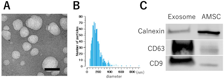
Hypoxic-ischemic brain injury arises from inadequate oxygen delivery to the brain, commonly occurring following cardiac arrest, which lacks effective treatments. Recent studies have demonstrated the therapeutic potential of exosomes released from mesenchymal stem cells. Given the challenge of systemic dilution associated with intravenous administration, intranasal delivery has emerged as a promising approach. In this study, we investigate the effects of intranasally administered exosomes in an animal model. Exosomes were isolated from the cell supernatants using the ultracentrifugation method. Brain injury was induced in Sprague-Dawley rats through a transient four-vessel occlusion model. Intranasal administration was conducted with 3 × 10⁸ exosome particles in 20 µL of PBS or PBS alone, administered daily for 7 days post-injury. Long-term cognitive behavioral assessments, biodistribution of exosomes, and histological evaluations of apoptosis and neuroinflammation were conducted. Exosomes were primarily detected in the olfactory bulb one hour after intranasal administration, subsequently distributing to the striatum and midbrain. Rats treated with exosomes exhibited substantial improvement in cognitive function up to 28 days after the insult, and demonstrated significantly fewer apoptotic cells along with higher neuronal cell survival in the hippocampus. Exosomes were found to be taken up by microglia, leading to a decrease in the expression of cytotoxic inflammatory markers.

Keywords: exosome; hypoxic-ischemic brain injury; inflammation; intranasal administration; mesenchymal stem cell.


Conflict of interest statement

Author, Sho Yamaguchi is employed by the company Kaneka. The remaining authors declare that the research was conducted in the absence of any commercial or financial relationships that could be construed as a potential conflict of interest.

Figures

Figure 1
Figure 1
Characteristics of exosome derived from AMSC. (A) Electron microscopy depicts exosomes with a rounded morphology and an approximate diameter of 100 nm. (B) Size distribution, ascertained by the nanoparticle analyzer, delineates exosome dimensions ranging from 70–400 nm, which showed 110 nm peak. (C) Western blot detection shows heightened expression of CD9 and CD63 in the exosome fraction, while a reduced signal is observed for Calnexin compared to the AMSC sample. Bar: 100 nm.
Figure 2
Figure 2
Short- and long-term memory impairment test. The animals that received intranasal exosomes exhibited better memory function in accuracy (correct arm entry/all arm entry), travel distance, reference error (wrong arm entry), and working error (entry into arms where pellets were previously collected). Black: exosome group, gray: PBS group, *: p < 0.05.
Figure 3
Figure 3
Biodistribution of exosome. A large number of exosomal signals (approx. 250 signals/mm2) can be found in the olfactory nerve starting from one hour after transplantation. The signal can also be found in the olfactory tract, striatum, and midbrain. Note that the Y axis is different between the olfactory nerve and others. The representative figure shows the signal at each area 24 h after exosome administration (bar = 20 μm).
Figure 4
Figure 4
Neural damage in hippocampus. (A) Apoptosis in hippocampus was evaluated in CA1 lesion using TUNEL staining at day 3. Apoptotic cells were significantly decreased in the exosome group compared with that of PBS group. (B) Neural degeneration was observed in CA1 lesion using Fluoro-Jade C staining at day 7. Exosome group exhibited significantly less degenerated neural cells compared with PBS group. Bar = 100 μm, *: p < 0.05.
Figure 5
Figure 5
Neural survival in hippocampus. Neural cells were evaluated with Nissl staining. Nuclear aggregation and structural alterations were observed in the PBS group, whereas these were preserved in the exosome group at both day 14 (A) and day 28 (B). Bar = 100 μm, *: p < 0.05, **: p < 0.01.
Figure 6
Figure 6
Microglial activation in hippocampus. Activated microglia were prominently observed in the PBS group, characterized by cell proliferation and a change in morphology to an amoeboid shape. In contrast, these changes were notably attenuated in the exosome-treated group (upper and left lower panels) Bar = 100 μm. Exosomes labeled with ExoSparkler (green) were found to co-localize with microglia (orange), suggesting the anti-inflammatory role of exosomes (Nuclear staning by DAPI. right lower panel). Bar = 20 μm, **: p < 0.01.
Figure 7
Figure 7
The exosome-treated group exhibited lower cytokine expression compared to the PBS-treated group. Levels of IL-1b (upper panel), IL-6 (middle panel), and TNF-α (lower panel) are depicted. Bar = 100 μm, *: p < 0.05.
Figure 8
Figure 8
In vitro analysis of anti-inflammatory effect of exosomes. Exosomes were administered into mouse BV2 cell. Cell viability (A), IL-6 production (B), and TNF-α production (C) were assessed between LPS group and LPS with exosome group. *: p < 0.05, **: p < 0.01.

References

    1. Hosseini M., Wilson R.H., Crouzet C., Amirhekmat A., Wei K.S., Akbari Y. Resuscitating the Globally Ischemic Brain: Ttm and Beyond. Neurotherapeutics. 2020;17:539–562. doi: 10.1007/s13311-020-00856-z. - DOI - PMC - PubMed
    1. Geocadin R.G., Wijdicks E., Armstrong M.J., Damian M., Mayer S.A., Ornato J.P., Rabinstein A., Suarez J.I., Torbey M.T., Dubinsky R.M., et al. Practice Guideline Summary: Reducing Brain Injury Following Cardiopulmonary Resuscitation: Report of the Guideline Development, Dissemination, and Implementation Subcommittee of the American Academy of Neurology. Neurology. 2017;88:2141–2149. doi: 10.1212/WNL.0000000000003966. - DOI - PMC - PubMed
    1. Perkins G.D., Callaway C.W., Haywood K., Neumar R.W., Lilja G., Rowland M.J., Sawyer K.N., Skrifvars M.B., Nolan J.P. Brain Injury after Cardiac Arrest. Lancet. 2021;398:1269–1278. doi: 10.1016/S0140-6736(21)00953-3. - DOI - PubMed
    1. Liu X., Jia X. Neuroprotection of Stem Cells against Ischemic Brain Injury: From Bench to Clinic. Transl. Stroke Res. 2023 doi: 10.1007/s12975-023-01163-3. - DOI - PMC - PubMed
    1. Kosaka N., Iguchi H., Ochiya T. Circulating Microrna in Body Fluid: A New Potential Biomarker for Cancer Diagnosis and Prognosis. Cancer Sci. 2010;101:2087–2092. doi: 10.1111/j.1349-7006.2010.01650.x. - DOI - PMC - PubMed

LinkOut - more resources