

Fabrication of Quercetin-Functionalized Morpholine and Pyridine Motifs-Laden Silk Fibroin Nanofibers for Effective Wound Healing in Preclinical Study
- PMID: 38675123
- PMCID: PMC11054860
- DOI: 10.3390/pharmaceutics16040462
Fabrication of Quercetin-Functionalized Morpholine and Pyridine Motifs-Laden Silk Fibroin Nanofibers for Effective Wound Healing in Preclinical Study
Abstract
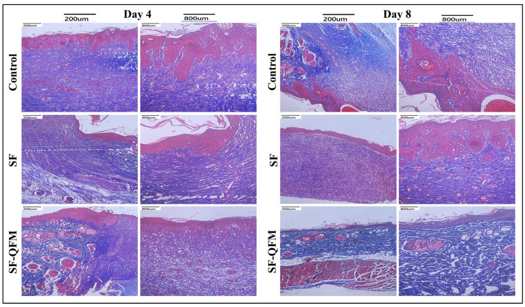
Choosing suitable wound dressings is crucial for effective wound healing. Spun scaffolds with bioactive molecule functionalization are gaining attention as a promising approach to expedite tissue repair and regeneration. Here, we present the synthesis of novel multifunctional quercetin with morpholine and pyridine functional motifs (QFM) embedded in silk fibroin (SF)-spun fibers (SF-QFM) for preclinical skin repair therapies. The verification of the novel QFM structural arrangement was characterized using ATR-FTIR, NMR, and ESI-MS spectroscopy analysis. Extensive characterization of the spun SF-QFM fibrous mats revealed their excellent antibacterial and antioxidant properties, biocompatibility, biodegradability, and remarkable mechanical and controlled drug release capabilities. SF-QFM mats were studied for drug release in pH 7.4 PBS over 72 h. The QFM-controlled release is mainly driven by diffusion and follows Fickian's law. Significant QFM release (40%) occurred within the first 6 h, with a total release of 79% at the end of 72 h, which is considered beneficial in effectively reducing bacterial load and helping expedite the healing process. Interestingly, the SF-QFM-spun mat demonstrated significantly improved NIH 3T3 cell proliferation and migration compared to the pure SF mat, as evidenced by the complete migration of NIH 3T3 cells within 24 h in the scratch assay. Furthermore, the in vivo outcome of SF-QFM was demonstrated by the regeneration of fresh fibroblasts and the realignment of collagen fibers deposition at 9 days post-operation in a preclinical rat full-thickness skin defect model. Our findings collectively indicate that the SF-QFM electrospun nanofiber scaffolds hold significant capability as a cost-effective and efficient bioactive spun architecture for use in wound healing applications.
Keywords: antibacterial; antioxidant; functionalized quercetin; rapid wound healing; spun silk fibroin; tissue engineering.
Conflict of interest statement
The authors declare no conflicts of interest.
Figures


References
-
- Dandona R., Kumar G.A., Gururaj G., James S., Chakma J.K., Thakur J.S., Srivastava A., Kumaresh G., Glenn S.D., Gupta G., et al. Mortality Due to Road Injuries in the States of India: The Global Burden of Disease Study 1990–2017. Lancet Public Health. 2020;5:e86–e98. doi: 10.1016/S2468-2667(19)30246-4. - DOI - PMC - PubMed
-
- Rezvani Ghomi E., Khalili S., Nouri Khorasani S., Esmaeely Neisiany R., Ramakrishna S. Wound Dressings: Current Advances and Future Directions. J. Appl. Polym. Sci. 2019;136:47738. doi: 10.1002/app.47738. - DOI
-
- Bolívar-Monsalve E.J., Alvarez M.M., Hosseini S., Espinosa-Hernandez M.A., Ceballos-González C.F., Sanchez-Dominguez M., Shin S.R., Cecen B., Hassan S., Di Maio E., et al. Engineering Bioactive Synthetic Polymers for Biomedical Applications: A Review with Emphasis on Tissue Engineering and Controlled Release. Mater. Adv. 2021;2:4447–4478. doi: 10.1039/D1MA00092F. - DOI
LinkOut - more resources
Full Text Sources
Molecular Biology Databases
Miscellaneous
