. 2024 Apr 1;16(4):485.
doi: 10.3390/pharmaceutics16040485.

Vanillin Promotes Osteoblast Differentiation, Mineral Apposition, and Antioxidant Effects in Pre-Osteoblasts

Affiliations

Vanillin Promotes Osteoblast Differentiation, Mineral Apposition, and Antioxidant Effects in Pre-Osteoblasts

Hyung-Mun Yun et al. Pharmaceutics. .

Abstract

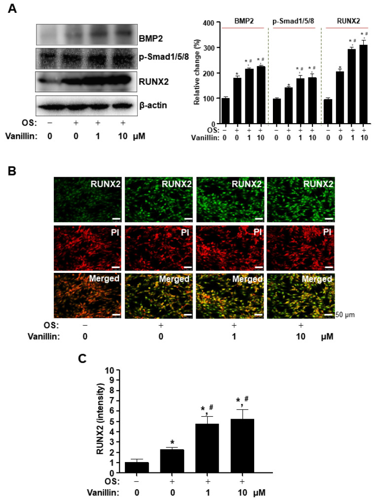
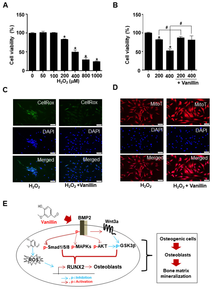
Antioxidant vanillin (4-hydroxy-3-methoxybenzaldehyde) is used as a flavoring in foods, beverages, and pharmaceuticals. Vanillin possesses various biological effects, such as antioxidant, anti-inflammatory, antibacterial, and anticancer properties. This study aimed to investigate the biological activities of vanillin purified from Adenophora triphylla var. japonica Hara on bone-forming processes. Vanillin treatment induced mineralization as a marker for mature osteoblasts, after stimulating alkaline phosphatase (ALP) staining and activity. The bone-forming processes of vanillin are mainly mediated by the upregulation of the bone morphogenetic protein 2 (BMP2), phospho-Smad1/5/8, and runt-related transcription factor 2 (RUNX2) pathway during the differentiation of osteogenic cells. Moreover, vanillin promoted osteoblast-mediated bone-forming phenotypes by inducing migration and F-actin polymerization. Furthermore, we validated that vanillin-mediated bone-forming processes were attenuated by noggin and DKK1. Finally, we demonstrated that vanillin-mediated antioxidant effects prevent the death of osteoblasts during bone-forming processes. Overall, vanillin has bone-forming properties through the BMP2-mediated biological mechanism, indicating it as a bone-protective compound for bone health and bone diseases such as periodontitis and osteoporosis.

Keywords: BMP2; Cbfa1; ROS; RUNX2; bone; mineralization; osteoblast; osteogenesis; vanillin.


Conflict of interest statement

The authors declare no conflict of interest.

Figures

Figure 1
Figure 1
Purification procedure, characterization, and structure of vanillin isolated from Adenophora triphylla var. japonica Hara. (A) Strategy for the isolation of vanillin. (B,C) 13C NMR (100 MHz, CD3OD) (B) and 1H NMR (400 MHz, CD3OD) (C) spectra obtained using JEOL ECX-500 spectrometer. (D) HPLC and chemical structure of vanillin (C8H8O3, Purity: >99.99%).
Figure 2
Figure 2
Effects of vanillin on cytotoxicity and osteoblast differentiation in osteogenic cells. (A) Cell toxicity was determined in osteogenic cells using the MTT assay. (BE) Cells were treated in osteogenic supplement medium (OS) containing 50 μg/mL L-ascorbic acid and 10 mM β-glycerophosphate with vanillin for 7 days (B,C) and 14 days (D,E). Osteoblast differentiation was analyzed by ALP staining (B), ALP activity (C), and ARS staining (D). ARS stains were eluted, and the ARS staining level was measured at 590 nm (E). Data are expressed as the mean ± SD. * p < 0.05, versus control. # p < 0.05, versus OS. Data represent the results of three experiments.
Figure 3
Figure 3
Effects of vanillin on the activation of BMP2 signaling in osteoblasts. (AC) Cells were treated in OS with vanillin for 3 days. Western blot analysis was performed to investigate the expression of BMP2, p-Smad1/5/8, RUNX2, and β-actin (A). RUNX2 was immunostained with rabbit anti-RUNX2 antibody, followed by Alex488-conjugated secondary antibody (green). And then, the cells were stained with PI (red). Images shown in the upper and middle panels were observed using multiphoton microscopy, and the images are merged in the lower bottom panels (B). The relative intensity is shown as a bar graph (C). Scale bar: 50 μm. Data are expressed as the mean ± SD. * p < 0.05, versus control. # p < 0.05, versus OS. Data represent the results of three experiments.
Figure 4
Figure 4
Effect of vanillin on BMP2-related signaling in osteoblasts. (AC) Cells were treated in OS with vanillin for 3 days. Western blot analysis was performed to investigate the expression of ERK, p-ERK, JNK, p-JNK, p38, p-p38, and β-actin (A); AKT, p-AKT, and β-actin (B); Wnt3a, GSK3β, p-GSK3β, and β-actin. The relative change (%) is shown as a bar graph. Data are expressed as the mean ± SD. * p < 0.05, versus control. # p < 0.05, versus OS. Data represent the results of three experiments.
Figure 5
Figure 5
Effects of vanillin on F-actin polymerization, cell migration, and the inhibition of BMP signaling in vanillin-stimulated osteoblast differentiation. (A) F-actin polymerization was stained with Fluorescein phalloidin (green), and the cells were stained with DRAQ5 (red). Images shown in the upper and middle panels were observed using multiphoton microscopy, and the images are merged in the lower bottom panels. Scale bar: 50 μm. (B) Cell migration was detected using a Matrigel-coated membrane in a Boyden chamber. (C,D) Vanillin was treated with noggin (10 μg/mL) or DKK1 (0.5 μg/mL) with OS for 7 days (C) and 14 days (D). Osteoblast differentiation was analyzed by ALP activity (C) and ARS staining (D). Data are expressed as the mean ± SD. * p < 0.05, versus control. # p < 0.05, versus OS. & p < 0.05, versus vanillin. Data represent the results of three experiments.
Figure 6
Figure 6
Antioxidant effects on oxidative stress of vanillin in osteoblasts. (A) Cells were treated in OS with indicated concentration of H2O2, and cell viability was determined using MTT assay. (B) Oxidative stress-induced cells in OS were treated with vanillin, and cell viability was determined using MTT assay. (C,D) ROS level (C) and active mitochondria (D) in oxidative stress-induced cells were detected using CellROX™ Green reagent and MitoTracker™ Red CMXRos, respectively. Scale bar: 50 μm. (E) Proposed model underlying antioxidant vanillin as protective compound in osteoblast differentiation and survival. Data are expressed as the mean ± SD. * p < 0.05, versus control. # p < 0.05, versus vanillin. Data represent the results of three experiments.

References

    1. Fakhry M., Hamade E., Badran B., Buchet R., Magne D. Molecular mechanisms of mesenchymal stem cell differentiation towards osteoblasts. World J. Stem Cells. 2013;5:136–148. doi: 10.4252/wjsc.v5.i4.136. - DOI - PMC - PubMed
    1. Phan T.C., Xu J., Zheng M.H. Interaction between osteoblast and osteoclast: Impact in bone disease. Histol. Histopathol. 2004;19:1325–1344. doi: 10.14670/HH-19.1325. - DOI - PubMed
    1. Park K.R., Lee J.Y., Kim B.M., Kang S.W., Yun H.M. TMARg, a Novel Anthraquinone Isolated from Rubia cordifolia Nakai, Increases Osteogenesis and Mineralization through BMP2 and beta-Catenin Signaling. Int. J. Mol. Sci. 2020;21:5332. doi: 10.3390/ijms21155332. - DOI - PMC - PubMed
    1. Lee H.R., Kim H.M., Jeong H.W., Kim G.G., Na C.I., Oh M.M., Hwang S.J. Growth Characteristics of Adenophora triphylla var. japonica Hara Seedlings as Affected by Growing Medium. Plants. 2019;8:466. doi: 10.3390/plants8110466. - DOI - PMC - PubMed
    1. Mlynarczyk K., Walkowiak-Tomczak D., Lysiak G.P. Bioactive properties of Sambucus nigra L. as a functional ingredient for food and pharmaceutical industry. J. Funct. Foods. 2018;40:377–390. doi: 10.1016/j.jff.2017.11.025. - DOI - PMC - PubMed

LinkOut - more resources