Review
. 2024 Apr 11;16(4):528.
doi: 10.3390/pharmaceutics16040528.

Mirk/Dyrk1B Kinase Inhibitors in Targeted Cancer Therapy

Affiliations
Review

Mirk/Dyrk1B Kinase Inhibitors in Targeted Cancer Therapy

Nikolaos Kokkorakis et al. Pharmaceutics. .

Abstract

During the last years, there has been an increased effort in the discovery of selective and potent kinase inhibitors for targeted cancer therapy. Kinase inhibitors exhibit less toxicity compared to conventional chemotherapy, and several have entered the market. Mirk/Dyrk1B kinase is a promising pharmacological target in cancer since it is overexpressed in many tumors, and its overexpression is correlated with patients' poor prognosis. Mirk/Dyrk1B acts as a negative cell cycle regulator, maintaining the survival of quiescent cancer cells and conferring their resistance to chemotherapies. Many studies have demonstrated the valuable therapeutic effect of Mirk/Dyrk1B inhibitors in cancer cell lines, mouse xenografts, and patient-derived 3D-organoids, providing a perspective for entering clinical trials. Since the majority of Mirk/Dyrk1B inhibitors target the highly conserved ATP-binding site, they exhibit off-target effects with other kinases, especially with the highly similar Dyrk1A. In this review, apart from summarizing the data establishing Dyrk1B as a therapeutic target in cancer, we highlight the most potent Mirk/Dyrk1B inhibitors recently reported. We also discuss the limitations and perspectives for the structure-based design of Mirk/Dyrk1B potent and highly selective inhibitors based on the accumulated structural data of Dyrk1A and the recent crystal structure of Dyrk1B with AZ191 inhibitor.

Keywords: Mirk/Dyrk1B kinase; X-ray crystal structures; apoptosis; cancer; cancer stem cells; inhibitors; quiescence; targeted cancer therapy.


Conflict of interest statement

The authors declare no conflicts of interest.

Figures

Figure 1
Figure 1
Summary of the major known functions of Dyrk1B. Dual-specificity tyrosine-regulated kinase 1B (Dyrk1B) plays a critical role in many biological processes in development and human disease, by regulating cell cycle progression/exit and quiescence, stemness maintenance, transcriptional regulation, cell motility, as well as cell survival, oxidative stress, and apoptosis.
Figure 2
Figure 2
Cross-talk of Dyrk1B in signaling pathways in development and disease. Serum mitogens down-regulate Dyrk1B through the RAS/RAF/MEK/ERK signaling pathway. In turn, Dyrk1B is implicated in glucose homeostasis through the inhibition of the RAS–RAF–MEK pathway in metabolic syndrome which is accompanied by diabetes. In cancer, Dyrk1B is involved in a complex cross-talk with Hedgehog (Hh). Dyrk1B inhibits ‘canonical’ Hh signaling initiated by Smoothened, while it promotes ‘non-canonical’ Hh signaling and oncogenic Gli1 activity by promoting PI3K/mTOR/AKT signaling and PI3K–AKT-mediated stability of the Gli1 transcription factor. Conversely, activated AKT directly inhibits the expression of Dyrk1B. In cancer, oncogenic mutant RAS (KRAS) initiates the ‘non-canonical’ HH pathway through the activation of Dyrk1B, via an elusive mechanism, employing several RAS effectors such as RAF/MEK/ERK, PI3K/AKT, and RLF/RAL. Dashed lines represent indirect mechanisms and yellow star represents phosphorylation HH: Hedgehog; ERK: Extracellular signal-regulated kinases; SMO: Smoothened.
Figure 3
Figure 3
Chemical structures of the Mirk/Dyrk1B kinase inhibitors. Chemical structures of known Dyrk1B inhibitors that are referred to this review. Notably, the VER-239353 inhibitor (compound 34) that belongs to pyrrolopyrimidine inhibitors [79] is different from the compound 34 that belongs to 2-alkylpyrimidine inhibitors [80].
Figure 4
Figure 4
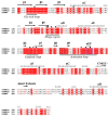
Sequence alignment of the kinase domains of human Dyrk1A (Uniprot: Q13627), Dyrk1B (Uniprot: Q9Y463), and Dyrk2 (Uniprot: Q92630) kinases. The main secondary structure elements and characteristic functional loops/regions are shown. The auto-phosphorylation site on the activation loops of Dyrk1A and Dyrk1Β is shown with an asterisk. Identical amino acid residues are highlighted in red and similar residues are shown in red letters. The Dyrk1A residues making critical (mostly polar) interactions with the bound ATP (see Section 4.2) are indicated with black boxes, whereas the residues interacting with a consensus peptide substrate (see Section 4.3) are indicated with inverted triangles. All of these residues are highly conserved in DYRKs, except Dyrk1A-Asn 244, which is replaced with an aspartic in Dyrk3 (Dyrk 3 and Dyrk 4 are not shown for simplicity). The conserved DFG motif of the activation loop is enclosed in a blue box and the conserved Arg residues interacting with the phosphorylated Tyr (shown with an asterisk) of the activation loop are enclosed in a black box (see Section 4.1). The sequence identity between kinase domains of Dyrk1A and Dyrk1B is 85%, whereas their identity with Dyrk2 is 45%.
Figure 5
Figure 5
Structural comparisons of activated and non-activated homologous kinases. (A) Superposition of Dyrk1A (green) with Dyrk2 (yellow). Dyrk1A (PDB: 2WO6) [98] is in complex with the consensus substrate peptide RARPTPALRE (in magenta) and with the ATP inhibitor DJM2005 (in orange), whereas Dyrk2 is in its Apo-form (PDB: 3K2L) [98]. The hinge region and the activation and catalytic loops of Dyrk1A are colored dark blue, light blue, and pink, respectively. (B) Superposition of the activated Dyrk1A (PDB: 2WO6; green) with the corresponding inactivated Abl kinase domain (PDB: 2HYY; light grey) [100]. The autophosphorylated Tyr 321 (p-Tyr 321) of Dyrk1A and the equivalent non-phosphorylated Tyr 393 of Abl (in blue italics) are shown as sticks. The section of the activation loop of Abl, colored in yellow, adopts the “inactive conformation”, occupying the substrate binding cleft. (C) Close-up views of the DFG-in and DFG-out motifs in Dyrk1A (PDB: 2WO6; green) and Abl (PDB: 2HYY; light grey), respectively. Dyrk1A residues are shown in black, whereas Abl residues are in blue italics. (D) Close-up view of the activation loop and of the substrate binding cleft of Dyrk1A (PDB: 2WO6). The polar interactions between p-Tyr 321 and Arg residues 325 and 328, locking the activation loop in its “active conformation”, are shown. Also shown is the DFG-in conformation of Dyrk1A, where Asp 307 together with Asn 292 can co-ordinate Mg2+ for the hydrolysis of the bound ATP molecule, and the orientation of the catalytic Lys 289 which catalyzes the phosphoryl transfer reaction from the ATP to the substrate. Images were created with PyMOL 1.8.0.4 version.
Figure 6
Figure 6
ATP- and substrate-binding sites of Dyrk1A. (A) Close-up view of ADPNP (ATP analogue) bound to the ATP-binding site of Dyrk1A (PDB: 7A4O) [79]. The polar interactions between Dyrk1A and ADPNP (stick representation) are only shown as dashed lines. (B) Close-up view of the interactions of the consensus substrate peptide RARPTPALRE with Dyrk1A (PDB: 2WO6) [98]. Polar interactions are shown as dashed lines. The Dyrk1A interacting residues are in black, whereas the residues of the peptide are in blue italics. The numbering of the peptide residues starts at 0 for the threonine residue which is due to phosphorylation. (C,D) Superposition of small (C) and large (D) ATP-competitive inhibitors with ADPNP as in their crystal structures with Dyrk1A. The adenosine moiety of ADPNP is in yellow and its phosphate groups are in orange. In panel (C), the natural inhibitor harmine is shown in green (PDB: 3ANR) [97] and the VER-239353 inhibitor in cyan (PDB: 7A5N) [79]. In panel (D), the AZ191 inhibitor is shown in light pink (PDB: 8C3G) [101] and the DJM2005 inhibitor (PDB: 2VX3) [98] in deep blue. Images were created with PyMOL 1.8.0.4 version.
Figure 7
Figure 7
Structural comparison of Dyk1A and Dyrk1B kinase domains. (A) Superposition of the AZ191-bound kinase domains of Dyrk1A (yellow; 8C3G) and Dyrk1B (green; 8C2Z) [101]. The bound AZ191 is shown in magenta or cyan for Dyrk1A or Dyrk1B, respectively. The phosphorylated tyrosine residues 321 or 273 of Dyrk1A or Dyrk1B, respectively, are shown in sticks on their activation loops. (B) Close-up view of important and identical polar interactions between AZ191 and Dyrk1A or Dyrk1B. The only difference in the ATP-binding site of these kinase domains is one residue in the hinge region (Dyrk1B-Leu 192 instead of Dyrk1A-Met 240). Images were created with PyMOL 1.8.0.4 version.

Similar articles

Cited by

References

    1. Dancey J., Sausville E.A. Issues and Progress with Protein Kinase Inhibitors for Cancer Treatment. Nat. Rev. Drug Discov. 2003;2:296–313. doi: 10.1038/nrd1066. - DOI - PubMed
    1. Arora A., Scholar E.M. Role of Tyrosine Kinase Inhibitors in Cancer Therapy. J. Pharmacol. Exp. Ther. 2005;315:971–979. doi: 10.1124/jpet.105.084145. - DOI - PubMed
    1. Grant S.K. Therapeutic Protein Kinase Inhibitors. Cell. Mol. Life Sci. 2009;66:1163–1177. doi: 10.1007/s00018-008-8539-7. - DOI - PMC - PubMed
    1. Cohen P., Cross D., Jänne P.A. Kinase Drug Discovery 20 Years after Imatinib. Nat. Rev. Drug Discov. 2022 doi: 10.1038/s41573-022-00418-2. - DOI - PubMed
    1. Magalhaes L.G., Ferreira L.L.G., Andricopulo A.D. Recent Advances and Perspectives in Cancer Drug Design. An. Acad. Bras. Ciências. 2018;90:1233–1250. doi: 10.1590/0001-3765201820170823. - DOI - PubMed

LinkOut - more resources