Review
. 2024 Apr 12;16(4):531.
doi: 10.3390/pharmaceutics16040531.

Cell Membrane-Coated Biomimetic Nanoparticles in Cancer Treatment

Affiliations
Review

Cell Membrane-Coated Biomimetic Nanoparticles in Cancer Treatment

Shu Zhang et al. Pharmaceutics. .

Abstract

Nanoparticle-based drug delivery systems hold promise for cancer treatment by enhancing the solubility and stability of anti-tumor drugs. Nonetheless, the challenges of inadequate targeting and limited biocompatibility persist. In recent years, cell membrane nano-biomimetic drug delivery systems have emerged as a focal point of research and development, due to their exceptional traits, including precise targeting, low toxicity, and good biocompatibility. This review outlines the categorization and advantages of cell membrane bionic nano-delivery systems, provides an introduction to preparation methods, and assesses their applications in cancer treatment, including chemotherapy, gene therapy, immunotherapy, photodynamic therapy, photothermal therapy, and combination therapy. Notably, the review delves into the challenges in the application of various cell membrane bionic nano-delivery systems and identifies opportunities for future advancement. Embracing cell membrane-coated biomimetic nanoparticles presents a novel and unparalleled avenue for personalized tumor therapy.

Keywords: biomimetic; cell membrane-coated nanoparticles; drug delivery; tumor targeting.


Conflict of interest statement

The authors declare no conflicts of interest.

Figures

Figure 1
Figure 1
The representative development and features of CMC@NPs (created by the authors using Adobe Illustrator 2023® software).
Figure 2
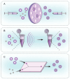
Figure 2
Preparation of CMC@NPs. (A) Co-extrusion. (B) Sonication. (C) Microfluidic electroporation (created by the authors using Adobe Illustrator 2023® software).
Figure 3
Figure 3
(A) The synthetic route for the lCUR-DOX@RBC. (B) Schematic illustration of chemo-immunotherapy of lCUR-DOX@RBC in tumor-bearing mice. Reprinted with permission from Ref. [31]. Copyright © 2024, Elsevier B.V: Amsterdam, The Netherlands.
Figure 4
Figure 4
Schematic of the thrombosis-mediated navigation system for P-aPD-1. Reprinted with permission from Ref. [43]. Copyright © 2024 The Authors, some rights reserved.
Figure 5
Figure 5
(A) In vivo living images after various treatments in tumor-excising mice. (a), PBS; (b), PPcDG/DiD; (c), NM/PPcDG/DiD. (B) In vivo imaging of tumors after tumor resection and at the end of therapy. (C) Typical tumor images in various groups at the end of therapy. (D) Representative pictures of lung tissues in various groups at the end of therapy (some metastatic nodules were in the back). (E) Full scanning of the H&E staining sections of the lung tissues in the PBS group and the NM/PPcDG/D group. Black circles indicated the typical metastasis in the PBS group (Scale bar: 1000 μm). Reprinted with permission from Ref. [7]. Copyright © 2024 Acta Materialia Inc.: Oxford, UK.
Figure 6
Figure 6
(A) Schematic illustration of (pMETTL14+RS09)@cRGD-M in dual-targeted tumor therapy. (B) Tumor volumes of different groups (** p < 0.01). (C,D) TUNEL staining indicated cell apoptosis in tumor tissues (Scale bar: 20 μm, ** p < 0.01). Reprinted with permission from Ref. [51]. Copyright © 2024 The Authors.
Figure 7
Figure 7
Schematic illustration of the TAM-membrane-coated upconversion NPs for improved photodynamic immunotherapy. (I) Preparation of NPR@TAMM. (II) Schematic representation of antitumor mechanism by NPR@TAMM. Reprinted with permission from Ref. [50]. Copyright © 2024 American Chemical Society: Washington, DC, USA.
Figure 8
Figure 8
Fabrication of N3-TINPs and their application for highly efficient photothermal therapy. Reprinted with permission from Ref. [57]. Copyright © 2024 The Authors.
Figure 9
Figure 9
(A,B) In vivo fluorescence imaging quantitative fluorescence intensity analysis after Cy5.5-labeled AIE NPs and CM@AIE NPs administrations, respectively (*** p < 0.001). (C) Immunofluorescence images of ZO-1 and CD31 on brain blood vessels after various treatments for 8 h (Scale bar: 100 μm). (D) H&E analysis of tumor slices after various treatments (Scale bar: 50 μm). Reprinted with permission from Ref. [56]. Copyright © 2024 Elsevier Ltd.: Oxford, UK.
Figure 10
Figure 10
Schematic illustration of the preparation of iDCs and the mechanism of synergy between iDCs and mild photothermal-immunotherapy. Reprinted with permission from Ref. [61]. Copyright © 2024 Elsevier Ltd.: Oxford, UK.
Figure 11
Figure 11
(A) Transwell schematic illustrations. (B) Confocal images. (C,D) Flow cytometric quantification of T and NK cells expressed by brain tumor tissue (**** p < 0.0001). (E) In vivo imaging system detection images and bioluminescence quantification of mice preimmunized with different formulas (** p < 0.01, *** p < 0.001). Reprinted with permission from Ref. [63]. Copyright © 2024 American Chemical Society: Washington, DC, USA.
Figure 12
Figure 12
(A) Intracellular free radical generation via DCFH-DA assay. (B) Live/dead cell staining via calcein-AM/PI assay. (C,D) Apoptosis assay via Annexin V-FITC/PI staining. Reprinted with permission from Ref. [65]. Copyright © 2024 The Authors.
Figure 13
Figure 13
(A) Schematic illustration of C-Z@CM multi-mode anti-tumor actions and the main process and chemical equation of ONOO production. (B) Representative ROS and ONOO staining images of tumor tissues after different treatments for 24 h (Scale bar: 100 μm). (C) Quantitative analyses of NO content in tumor tissue (** p < 0.01). Reprinted with permission from Ref. [74]. Copyright © 2024 Wiley-VCH GmbH.
Figure 14
Figure 14
(A) Scheme of CMO-R@4T1-mediated targeted NIR-II photothermal immunotherapy. (B) In vivo photoacoustic imaging and intensity of tumor-bearing mice with the tumor regions highlighted (yellow circle) (Scale bar: 5 mm). (C,D) Tumor growth curves of primary and distant tumors on bilateral tumor-bearing mice after various treatments (*** p < 0.001). (E) Immunofluorescence staining of CD8+ T cells (green) and CD4+ T cells (red) from both primary and distant tumor tissues of mice after different treatments (Scale bar: 100 μm). Reprinted with permission from Ref. [77]. Copyright © 2024, Wiley-VCH: Weinheim, Germany.
Figure 15
Figure 15
(A) In vivo real-time fluorescence images (Scale bar: 20 μm). (B) In vitro fluorescence images of tumors and major organs. (C) Infrared thermography of tumor-bearing mice. (D) H&E staining of the tumor sections (I: Saline, II: Fe3O4, III: R837, IV: ICG, V: RFe, VI: RIFe + NIR, VII: RIFe@TRM, VIII: RIFe@TRM + NIR, IX: RIFe@TRM + NIR + MF, Scale bar: 20 μm). Reprinted with permission from Ref. [86]. Copyright © 2024 American Chemical Society: Washington, DC, USA.
Figure 16
Figure 16
Design of tumor microenvironment targeting for glioblastoma multiforme treatment via hybrid cell membrane coating supramolecular micelles. Reprinted with permission from Ref. [90]. Copyright © 2024 Elsevier B.V: Amsterdam, The Netherlands.
Figure 17
Figure 17
Different classification of CMC@NPs used for cancer therapy (Created by authors using Adobe Illustrator 2023® software).

Similar articles

Cited by

References

    1. Siegel R.L., Miller K.D., Wagle N.S., Jemal A. Cancer statistics, 2023. CA Cancer J Clin. 2023;73:17–48. doi: 10.3322/caac.21763. - DOI - PubMed
    1. Desai N. Challenges in development of nanoparticle-based therapeutics. AAPS J. 2012;14:282–295. doi: 10.1208/s12248-012-9339-4. - DOI - PMC - PubMed
    1. Xu P., Zuo H., Chen B., Wang R., Ahmed A., Hu Y., Ouyang J. Doxorubicin-loaded platelets as a smart drug delivery system: An improved therapy for lymphoma. Sci. Rep. 2017;7:42632. doi: 10.1038/srep42632. - DOI - PMC - PubMed
    1. Lubich C., Allacher P., Maurus D.L.R., Bauer A., Prenninger T., Horling F.M., Siekmann J., Oldenburg J., Scheiflinger F., Reipert B.M. The Mystery of Antibodies Against Polyethylene Glycol (PEG)—What do we Know? Pharm. Res. 2016;33:2239–2249. doi: 10.1007/s11095-016-1961-x. - DOI - PubMed
    1. Hwang J., Jeong Y., Park J.M., Lee K.H., Hong J.W., Choi J. Biomimetics: Forecasting the future of science, engineering, and medicine. Int. J. Nanomed. 2015;10:5701–5713. - PMC - PubMed

LinkOut - more resources