. 2024 Apr 11;29(8):1737.
doi: 10.3390/molecules29081737.

A Novel Non-Psychoactive Fatty Acid from a Marine Snail, Conus inscriptus, Signals Cannabinoid Receptor 1 (CB1) to Accumulate Apoptotic C16:0 and C18:0 Ceramides in Teratocarcinoma Cell Line PA1

Affiliations

A Novel Non-Psychoactive Fatty Acid from a Marine Snail, Conus inscriptus, Signals Cannabinoid Receptor 1 (CB1) to Accumulate Apoptotic C16:0 and C18:0 Ceramides in Teratocarcinoma Cell Line PA1

Christina Sathyanathan Vijayaraghavan et al. Molecules. .

Abstract

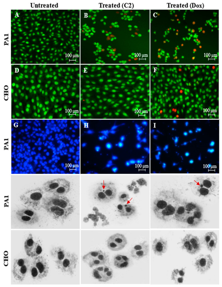
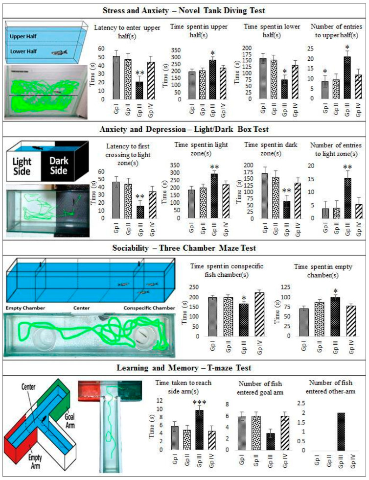
The cannabinoid-type I (CB1) receptor functions as a double-edged sword to decide cell fate: apoptosis/survival. Elevated CB1 receptor expression is shown to cause acute ceramide accumulation to meet the energy requirements of fast-growing cancers. However, the flip side of continual CB1 activation is the initiation of a second ceramide peak that leads to cell death. In this study, we used ovarian cancer cells, PA1, which expressed CB1, which increased threefold when treated with a natural compound, bis(palmitoleic acid) ester of a glycerol (C2). This novel compound is isolated from a marine snail, Conus inscriptus, using hexane and the structural details are available in the public domain PubChem database (ID: 14275348). The compound induced two acute ceramide pools to cause G0/G1 arrest and killed cells by apoptosis. The compound increased intracellular ceramides (C:16 to 7 times and C:18 to 10 times), both of which are apoptotic inducers in response to CB1 signaling and thus the compound is a potent CB1 agonist. The compound is not genotoxic because it did not induce micronuclei formation in non-cancerous Chinese hamster ovarian (CHO) cells. Since the compound induced the cannabinoid pathway, we tested if there was a psychotropic effect in zebrafish models, however, it was evident that there were no observable neurobehavioral changes in the treatment groups. With the available data, we propose that this marine compound is safe to be used in non-cancerous cells as well as zebrafish. Thus, this anticancer compound is non-toxic and triggers the CB1 pathway without causing psychotropic effects.

Keywords: FAAH1; MD simulation; apoptosis; cannabinoid receptor 1 (CB1); ceramide; ovarian cancer; zebrafish models.


Conflict of interest statement

The authors declare no potential conflicts of interest.

Figures

Figure 1
Figure 1
PA1 cell lines were treated with C2 (IC50 1 µg/mL (1.7 µM)) and a reference drug, doxorubicin (IC50 5 µg/mL (8.6 µM)). Acridine orange/propidium iodide-stained (AF) and DAPI-stained (GI) cells in epifluorescent microscope view show apoptotic bodies (red), while the CHO cells were found healthy upon C2 treatment. Micronuclei formation (red arrows) was seen for C2- and doxorubicin-treated PA1 cells at 20× in an Axioscope A1 Biology Microscope which is not visible for CHO, indicating C2 is not genotoxic in non-cancerous cell lines.
Figure 2
Figure 2
C2/Doxorubicin-treated (A) PA1 cell migration was inhibited (gap filling compared for 0 and 24 h and the percentages are given at 24 h: 1.09 and 2.4%, respectively) compared to their untreated controls (86.4%), showing their anti-invasive properties (B) [* p < 0.05, ** p ˂ 0.005]. The cells accumulated at G0/G1 phase for the C2-treated groups (D) and at G2/M for doxorubicin (E) compared to their untreated controls where cells were distributed across all phases (C) (color code: green—G0; red—G0/G1; blue—S; and pink—G2/M phase). Clear MMP depolarization was seen in C2-treated PA1 cells (75.6%) (G) from the untreated controls (8.1%) (F). (E,H) correspond to doxorubicin-treated positive controls.
Figure 3
Figure 3
Enhanced expression of mitochondrial-associated (BAX and BAD) and death receptor (FasL and FADDR) transcripts alongside restricted antiapoptotic (Bcl-xL, Bcl-2 and MMP2) mRNA transcripts (on a real-time basis), hinting at intrinsic as well as extrinsic apoptotic routes for C2 (* p ˂ 0.05, ** p ˂ 0.005, *** p ˂ 0.001) in PA1 cells. In silico analysis predicted that C2 binds to CB1 receptor as well as FAAH1, a membrane-bound enzyme (which degrades endogenous anandamide), as confirmed by molecular docking, MD simulations and RMSD results (Figure S2). CB1 expression both at gene (threefold higher) and protein levels (immunoblots are provided; β-actin served as loading control) was triggered upon treatment with C2. On the contrary, FAAH1 expression was 0.4 times lower than that of untreated control and this could be a reason for the maintenance of steady bioavailability of C2 without degradation in the treated PA1 cell population.
Figure 4
Figure 4
Ceramide peaks at C16:0 and C18:0 respectively were seven (B) and ten times (D) higher in the treated over control groups ((A) and (C) respectively) at the end of 16 h treatment regimen emphasizing C2-induced over-production of ceramides.
Figure 5
Figure 5
Behavioral effects of C2 using zebrafish models: Evaluation of anxiolytic/anxiogenic properties of C2 using novel tank diving test, light/dark box test and three-chamber maze test. All three tests revealed that both high and mid-doses are anxiolytic and that C2 treatment does not alter neuro-related functions though C2 is a ligand of CB1. Note that the ethanol control groups had anxiety-specific endpoints, like inhibition to diving, longer time spent in the upper half of the tank/bright zones/empty chamber. C2 is also shown to not affect the learning and memory capabilities of the fish as seen in the T-maze test. Statistically significant differences between C2- and ethanol-treated groups are indicated as * p < 0.05, ** p ˂ 0.005, *** p ˂ 0.001. Group I—high dose C2 (100 mg); Group II—mid-dose C2 (75 mg); Group III—ethanol control and Group IV—untreated control. Each group contained six fish and the results are presented as average values ± SE. Panels are based on our own observations validated using MATLAB-based tracking software (Mathworks Inc., Natick, MA, USA).
Figure 6
Figure 6
Representative hematoxylin and eosin (H,E)-stained photomicrographs showing sagittal section of hind brain tissues of zebrafish provide evidence of ventricle (A,B) and olfactory lobes (C,D) with normal architecture. The sagittal sections of cerebellum across all the groups show normal architecture (EH) indicative of (at least) no acute brain damage upon treatments with C2 (high and mid-doses) (long term exposures need to be ascertained).

Similar articles

References

    1. Hannun Y.A. Functions of Ceramide in Coordinating Cellular Responses to Stress. Science. 1996;274:1855–1859. doi: 10.1126/science.274.5294.1855. - DOI - PubMed
    1. Westwick J., Bielawska A., Dbaibo G., Hannun Y.A., Brenner D.A. Ceramide Activates the Stress-Activated Protein Kinases. J. Biol. Chem. 1995;270:22689–22692. doi: 10.1074/jbc.270.39.22689. - DOI - PubMed
    1. Latinis K.M., Koretzky G.A. Fas Ligation Induces Apoptosis and Jun Kinase Activation Independently of CD45 and Lck in Human T Cells. Blood. 1996;87:871–875. doi: 10.1182/blood.V87.3.871.bloodjournal873871. - DOI - PubMed
    1. Jayadev S., Liu B., Bielawska A., Lee J.Y., Nazaire F., Pushkareva M.Y., Obeid L.M., Hannun Y.A. Role for Ceramide in Cell Cycle Arrest. J. Biol. Chem. 1995;270:2047–2052. doi: 10.1074/jbc.270.5.2047. - DOI - PubMed
    1. Dbaibo G. Regulation of the Stress Response by Ceramide. Biochem. Soc. 1997;25:557–561. doi: 10.1042/bst0250557. - DOI - PubMed