. 2024 Apr 19;12(4):441.
doi: 10.3390/vaccines12040441.

The Papain-like Protease Domain of Severe Acute Respiratory Syndrome Coronavirus 2 Conjugated with Human Beta-Defensin 2 and Co1 Induces Mucosal and Systemic Immune Responses against the Virus

Affiliations

The Papain-like Protease Domain of Severe Acute Respiratory Syndrome Coronavirus 2 Conjugated with Human Beta-Defensin 2 and Co1 Induces Mucosal and Systemic Immune Responses against the Virus

Byeol-Hee Cho et al. Vaccines (Basel). .

Abstract

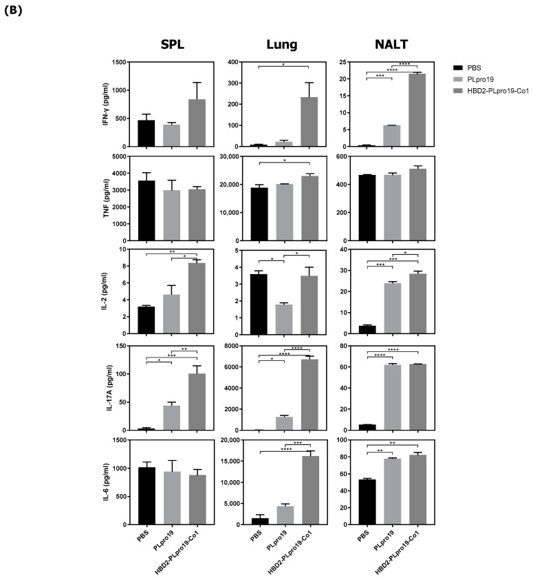
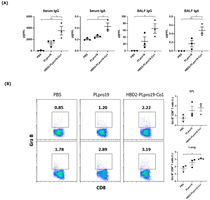
Most of the licensed vaccines against SARS-CoV-2 target spike proteins to induce viral neutralizing antibodies. However, currently prevalent SARS-CoV-2 variants contain many mutations, especially in their spike proteins. The development of vaccine antigens with conserved sequences that cross-react with variants of SARS-CoV-2 is needed to effectively defend against SARS-CoV-2 infection. Given that viral infection is initiated in the respiratory mucosa, strengthening the mucosal immune response would provide effective protection. We constructed a mucosal vaccine antigen using the papain-like protease (PLpro) domain of non-structural protein 3 of SARS-CoV-2. To potentiate the mucosal immune response, PLpro was combined with human beta-defensin 2, an antimicrobial peptide with mucosal immune adjuvant activity, and Co1, an M-cell-targeting ligand. Intranasal administration of the recombinant PLpro antigen conjugate into C57BL/6 and hACE2 knock-in (KI) mice induced antigen-specific T-cell and antibody responses with complement-dependent cytotoxic activity. Viral challenge experiments using the Wuhan and Delta strains of SARS-CoV-2 provided further evidence that immunized hACE2 KI mice were protected against viral challenge infections. Our study shows that PLpro is a useful candidate vaccine antigen against SARS-CoV-2 infection and that the inclusion of human beta-defensin 2 and Co1 in the recombinant construct may enhance the efficacy of the vaccine.

Keywords: SARS-CoV-2; adjuvant; papain-like protease; recombinant antigen; vaccine.


Conflict of interest statement

The authors declare no conflicts of interest.

Figures

Figure 1
Figure 1
Ag-specific Ab responses in C57BL/6 mice immunized with recombinant proteins. (A) Recombinant protein containing the sequence of the PLpro domain in non-structural protein 3 (Nsp3) of SARS-CoV-2. (B) Mice were immunized intranasally 5 times with 10 μg of PLpro19 or HBD2-PLpro19-Co1 once a week. Ag-specific IgG and IgA in the serum and BALF of immunized C57BL/6 were analyzed by ELISA 1 week after the last immunization. (C) After a 1 h incubation of infected Vero E6 with serum from mice, SARS-CoV-R+C3+ Vero E6 cells were detected by flow cytometry. (D) Subclasses of PLpro19-specific IgG were analyzed by ELISA. Data are presented as means ± standard errors (SEs) of repeated experiments and analyzed in a one-way ANOVA (* p < 0.05, ** p < 0.01, *** p < 0.001, **** p < 0.0001, n = 4). AC, acidic domain; X, X-domain; SUD, SARS-unique domain; PLpro, Papain-like protease; TM, transmembrane region; Y, Y-domain.
Figure 1
Figure 1
Ag-specific Ab responses in C57BL/6 mice immunized with recombinant proteins. (A) Recombinant protein containing the sequence of the PLpro domain in non-structural protein 3 (Nsp3) of SARS-CoV-2. (B) Mice were immunized intranasally 5 times with 10 μg of PLpro19 or HBD2-PLpro19-Co1 once a week. Ag-specific IgG and IgA in the serum and BALF of immunized C57BL/6 were analyzed by ELISA 1 week after the last immunization. (C) After a 1 h incubation of infected Vero E6 with serum from mice, SARS-CoV-R+C3+ Vero E6 cells were detected by flow cytometry. (D) Subclasses of PLpro19-specific IgG were analyzed by ELISA. Data are presented as means ± standard errors (SEs) of repeated experiments and analyzed in a one-way ANOVA (* p < 0.05, ** p < 0.01, *** p < 0.001, **** p < 0.0001, n = 4). AC, acidic domain; X, X-domain; SUD, SARS-unique domain; PLpro, Papain-like protease; TM, transmembrane region; Y, Y-domain.
Figure 2
Figure 2
Ag-specific T-cell immune responses in C57BL/6 mice immunized with recombinant proteins. (A) Lymphocytes prepared from the lung and splenocytes were incubated with PLpro19 protein (1 μg) for 24 h. Grz B+ CD8+ cells were analyzed by flow cytometry. (B) Cytokines were detected by cytometric bead array (CBA). The experiments were repeated three times and representative results are shown. All data were analyzed in a one-way ANOVA (* p < 0.05, ** p < 0.01, *** p < 0.001, **** p < 0.0001, n = 3). The results are presented as means ± SE, with the exception of NALT (mean ± standard deviation [SD]).
Figure 2
Figure 2
Ag-specific T-cell immune responses in C57BL/6 mice immunized with recombinant proteins. (A) Lymphocytes prepared from the lung and splenocytes were incubated with PLpro19 protein (1 μg) for 24 h. Grz B+ CD8+ cells were analyzed by flow cytometry. (B) Cytokines were detected by cytometric bead array (CBA). The experiments were repeated three times and representative results are shown. All data were analyzed in a one-way ANOVA (* p < 0.05, ** p < 0.01, *** p < 0.001, **** p < 0.0001, n = 3). The results are presented as means ± SE, with the exception of NALT (mean ± standard deviation [SD]).
Figure 3
Figure 3
Maintenance of Ag-specific immune responses in mice. (A) C57BL/6 mice were boosted with 10 μg of PLpro19 or HBD2-PLpro19-Co1 2 months after the last immunization. PLpro19-specific IgG and IgA in the serum and BALF of immunized mice were detected by ELISA 1 week after boosting. (B) Lymphocytes from lung and splenocytes were incubated with the PLpro19 protein (1 μg) for 24 h. Grz B+ CD8+ cells were analyzed by flow cytometry. (C) Cytokines were detected by CBA. (D) CD44hi CD4+ and CD8+ T-cells detected in the lung. The experiments were repeated twice and representative results are shown. The data were analyzed in a one-way ANOVA (* p < 0.05, ** p < 0.01, *** p < 0.001, **** p < 0.0001, n = 3) and are presented as means ± SE, with the exception of NALT (mean ± SD).
Figure 3
Figure 3
Maintenance of Ag-specific immune responses in mice. (A) C57BL/6 mice were boosted with 10 μg of PLpro19 or HBD2-PLpro19-Co1 2 months after the last immunization. PLpro19-specific IgG and IgA in the serum and BALF of immunized mice were detected by ELISA 1 week after boosting. (B) Lymphocytes from lung and splenocytes were incubated with the PLpro19 protein (1 μg) for 24 h. Grz B+ CD8+ cells were analyzed by flow cytometry. (C) Cytokines were detected by CBA. (D) CD44hi CD4+ and CD8+ T-cells detected in the lung. The experiments were repeated twice and representative results are shown. The data were analyzed in a one-way ANOVA (* p < 0.05, ** p < 0.01, *** p < 0.001, **** p < 0.0001, n = 3) and are presented as means ± SE, with the exception of NALT (mean ± SD).
Figure 4
Figure 4
Immunogenicity of PLpro19 constructs in hACE2 KI mice. (A) hACE2 KI mice were immunized intranasally five times with 10 μg of PLpro19 or HBD2-PLpro19-Co1 once a week. The levels of PLpro19-specific IgG and IgA in the serum and BALF of the immunized hACE2 KI mice were determined by ELISA 1 week after the last immunization. (B) Cells isolated from the spleen and lung were stimulated with PLpro19 protein (1 μg) for 24 h. Grz B+ CD8+ T-cells were analyzed by flow cytometry. (C) Cytokines were detected by CBA. The experiments were repeated three times and representative results are shown. All data were analyzed in a one-way ANOVA (* p < 0.05, ** p < 0.01, *** p < 0.001, **** p < 0.0001, n = 4) and are presented as means ± SE, with the exception of NALT (mean ± SD).
Figure 4
Figure 4
Immunogenicity of PLpro19 constructs in hACE2 KI mice. (A) hACE2 KI mice were immunized intranasally five times with 10 μg of PLpro19 or HBD2-PLpro19-Co1 once a week. The levels of PLpro19-specific IgG and IgA in the serum and BALF of the immunized hACE2 KI mice were determined by ELISA 1 week after the last immunization. (B) Cells isolated from the spleen and lung were stimulated with PLpro19 protein (1 μg) for 24 h. Grz B+ CD8+ T-cells were analyzed by flow cytometry. (C) Cytokines were detected by CBA. The experiments were repeated three times and representative results are shown. All data were analyzed in a one-way ANOVA (* p < 0.05, ** p < 0.01, *** p < 0.001, **** p < 0.0001, n = 4) and are presented as means ± SE, with the exception of NALT (mean ± SD).
Figure 5
Figure 5
Protective immune response against SARS-CoV-2 challenge infection. (A) hACE2 KI mice (n = 7) were immunized five times with 10 μg of PLpro19 or HBD2-PLpro19-Co1 once a week. A week after the last immunization, the mice were infected intranasally with SARS-CoV-2. The mice were weighed daily for 8 days after infection with the Wuhan strain of SARS-CoV-2 (2 × 10⁵ PFUs). (B) RNA expression of the S protein gene of SARS-CoV-2 in lung was detected by qRT-PCR 8 days post-infection (n = 2). (C) hACE2 KI mice (n = 6 for PBS, n = 7 for PLpro19 and HBD2-PLpro19-Co1) were weighed daily for 8 days after infection with the Delta strain of SARS-CoV-2 (1 × 10⁵ PFUs). (D) RNA expression of the N protein of SARS-CoV-2 in the lung was detected by qRT-PCR 8 days after infection (n = 2). The experiments were repeated twice and representative results are shown. The changes in body weight of the mice were analyzed in a two-way ANOVA, and the qRT-PCR data in a one-way ANOVA (* p < 0.05, ** p < 0.01, **** p < 0.0001). The data are presented as means ± SE.
Figure 6
Figure 6
PLpro19-specific immune responses 2 months after the immunization of hACE2 KI mice. (A) hACE2 mice were boosted with 10 μg of PLpro19 or HBD2-PLpro19-Co1 2 months after the last immunization. PLpro19-specific IgG and IgA in the serum and BALF of immunized hACE2 KI mice were detected by ELISA 1 week after boosting. (B) Cells isolated from the spleen and lung of hACE2 KI mice were stimulated with PLpro19 protein (1 μg) for 24 h. Grz B+ CD8+ T-cells were analyzed by flow cytometry. (C) CD44hi CD4+ and CD8+ T-cells were detected in the lung. The experiments were repeated three times and representative results are shown. All data were analyzed in a one-way ANOVA (* p < 0.05, ** p < 0.01, *** p < 0.001, **** p < 0.0001, n = 4). The data are presented as means ± SE.
Figure 6
Figure 6
PLpro19-specific immune responses 2 months after the immunization of hACE2 KI mice. (A) hACE2 mice were boosted with 10 μg of PLpro19 or HBD2-PLpro19-Co1 2 months after the last immunization. PLpro19-specific IgG and IgA in the serum and BALF of immunized hACE2 KI mice were detected by ELISA 1 week after boosting. (B) Cells isolated from the spleen and lung of hACE2 KI mice were stimulated with PLpro19 protein (1 μg) for 24 h. Grz B+ CD8+ T-cells were analyzed by flow cytometry. (C) CD44hi CD4+ and CD8+ T-cells were detected in the lung. The experiments were repeated three times and representative results are shown. All data were analyzed in a one-way ANOVA (* p < 0.05, ** p < 0.01, *** p < 0.001, **** p < 0.0001, n = 4). The data are presented as means ± SE.

Similar articles

References

    1. Chakraborty C., Bhattacharya M., Dhama K. SARS-CoV-2 vaccines, vaccine development technologies, and significant efforts in vaccine development during the pandemic: The lessons learned might help to fight against the next pandemic. Vaccines. 2023;11:682. doi: 10.3390/vaccines11030682. - DOI - PMC - PubMed
    1. Chen J., Xu W., Li L., Yi L., Jiang Y., Hao P., Xu Z., Zou W., Li P., Gao Z. Immunogenicity and protective potential of chimeric virus-like particles containing SARS-CoV-2 spike and H5N1 matrix 1 proteins. Front. Cell. Infect. Microbiol. 2022;12:967493. doi: 10.3389/fcimb.2022.967493. - DOI - PMC - PubMed
    1. Li L., Honda-Okubo Y., Huang Y., Jang H., Carlock M.A., Baldwin J., Piplani S., Bebin-Blackwell A.G., Forgacs D., Sakamoto K. Immunisation of ferrets and mice with recombinant SARS-CoV-2 spike protein formulated with Advax-SM adjuvant protects against COVID-19 infection. Vaccine. 2021;39:5940–5953. doi: 10.1016/j.vaccine.2021.07.087. - DOI - PMC - PubMed
    1. Zhang Z., Mateus J., Coelho C.H., Dan J.M., Moderbacher C.R., Gálvez R.I., Cortes F.H., Grifoni A., Tarke A., Chang J. Humoral and cellular immune memory to four COVID-19 vaccines. Cell. 2022;185:2434–2451.e17. doi: 10.1016/j.cell.2022.05.022. - DOI - PMC - PubMed
    1. Korber B., Fischer W.M., Gnanakaran S., Yoon H., Theiler J., Abfalterer W., Hengartner N., Giorgi E.E., Bhattacharya T., Foley B. Tracking changes in SARS-CoV-2 spike: Evidence that D614G increases infectivity of the COVID-19 virus. Cell. 2020;182:812–827.e19. doi: 10.1016/j.cell.2020.06.043. - DOI - PMC - PubMed

LinkOut - more resources