Review
. 2024 Mar 25;16(4):500.
doi: 10.3390/v16040500.

Friends and Foes: The Ambivalent Role of Autophagy in HIV-1 Infection

Affiliations
Review

Friends and Foes: The Ambivalent Role of Autophagy in HIV-1 Infection

Susanne Klute et al. Viruses. .

Abstract

Autophagy has emerged as an integral part of the antiviral innate immune defenses, targeting viruses or their components for lysosomal degradation. Thus, successful viruses, like pandemic human immunodeficiency virus 1 (HIV-1), evolved strategies to counteract or even exploit autophagy for efficient replication. Here, we provide an overview of the intricate interplay between autophagy and HIV-1. We discuss the impact of autophagy on HIV-1 replication and report in detail how HIV-1 manipulates autophagy in infected cells and beyond. We also highlight tissue and cell-type specifics in the interplay between autophagy and HIV-1. In addition, we weigh exogenous modulation of autophagy as a putative double-edged sword against HIV-1 and discuss potential implications for future antiretroviral therapy and curative approaches. Taken together, we consider both antiviral and proviral roles of autophagy to illustrate the ambivalent role of autophagy in HIV-1 pathogenesis and therapy.

Keywords: HIV; autophagy; innate immunity.


Conflict of interest statement

The authors declare no conflicts of interest.

Figures

Figure 1
Figure 1
Schematic overview of the autophagy pathway. (Left panel) Upon autophagy induction AMPK activates ULK1. Negative regulation is provided by the mTORC1/2 and Casein kinase II complexes. The activation of the PI3KC3 complex I promotes PI3P production at the endoplasmic reticulum. PI3P assembles WIPI proteins and DFCP1 to promote initial phagophore formation. To the phagophore, cytoplasmic cargo is recruited via selective autophagy receptors (SARs). LC3 is proteolytically cleaved by ATG4 to form LC3-I. A ubiquitin-like conjugation process mediated by ATG 7, ATG3 and ATG5-ATG12-ATG16L1 attaches phosphatidylethanolamine (PE) to LC3-I to generate LC3-II, which decorates the inner and outer membrane the phagophore. The phagophore matures into the double-membrane vesicle termed the autophagosome. Mediated by RAB proteins, SNARE proteins, the PI3KC3 complex II and the HOPS complex, the autophagosome fuses with a lysosome, forming the autophagolysosome and leading to the degradation of the cargo, inner membrane and SARs. Green arrows indicate positive stimulation; red arrows indicate negative regulation. (Right panel) Core proteins of the complexes involved in autophagy.
Figure 2
Figure 2
Schematic depiction of the genome of HIV-1. Group-specific antigens, gag. Polymerase, pol. Envelope, env. Viral infectivity factor, vif. Viral protein R, vpr. Viral protein U, vpu. Negative factor, nef. Regulator of expression of virion proteins, rev. Trans-activator of transcription, tat.
Figure 3
Figure 3
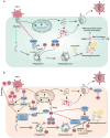
The interplay between autophagy and HIV-1 proteins. (a), Autophagy conveys the degradation of virions and viral proteins. For example, TRIM5α targets p24, Tat is degraded via p62 and HDAC6 targets the HIV-1 proteins Vif and p55 for autophagic clearance. Degradation of virions/viral components by autophagy provides PAMPs triggering PRR-dependent innate immune activation. Viral antigens are processed via autophagy and loaded on MHC-II molecules. (b), HIV-1 proteins such as Vpr, Tat and p17 trigger autophagy in infected cells. Env as well as Tat modulates autophagy in bystander cells. Tat induces autophagy via mediator PKM2-mTOR-AMPK. Vpr blocks FOXO3-mediated transcription of autophagy genes. Nef inhibits the pro-autophagic TFEB, promotes activation of BCL2, targets HDAC6 for degradation, inactivates the PI3KC3 complex II and interferes with STX17-mediated fusion of autophagosomes with lysosomes. Gag associates with LC3-II to assist its processing. Vpr triggers the degradation of SNAPIN. Tat inhibits autophagic maturation by interacting with LAMP2. Vpu mediates Tetherin restriction and promotes HIV-1 budding. Green arrows indicate positive stimulation; red arrows indicate negative regulation; Red crosses indicate inhibition.
Figure 4
Figure 4
The proposed cell-type-specific role of autophagy in HIV-1 infection. In CD4+ T cells, infection initiates a mainly antiviral autophagy response, which contributes to cell death of the infected cells. Released Env triggers autophagy in bystander CD4+ T cells, causing apoptosis. In infected macrophages, autophagy is activated and promotes HIV-1 replication. Secreted Tat modulates autophagy in bystander macrophages.

References

    1. Klionsky D.J. Autophagy: From Phenomenology to Molecular Understanding in Less than a Decade. Nat. Rev. Mol. Cell Biol. 2007;8:931–937. doi: 10.1038/nrm2245. - DOI - PubMed
    1. He C., Klionsky D.J. Regulation Mechanisms and Signaling Pathways of Autophagy. Annu. Rev. Genet. 2009;43:67–93. doi: 10.1146/annurev-genet-102808-114910. - DOI - PMC - PubMed
    1. Yorimitsu T., Klionsky D.J. Autophagy: Molecular Machinery for Self-Eating. Cell Death Differ. 2005;12:1542–1552. doi: 10.1038/sj.cdd.4401765. - DOI - PMC - PubMed
    1. Feng Y., He D., Yao Z., Klionsky D.J. The Machinery of Macroautophagy. Cell Res. 2014;24:24–41. doi: 10.1038/cr.2013.168. - DOI - PMC - PubMed
    1. Levine B., Mizushima N., Virgin H.W. Autophagy in Immunity and Inflammation. Nature. 2011;469:323–335. doi: 10.1038/nature09782. - DOI - PMC - PubMed

Publication types