

Sudden Infant Death Associated with Rhinovirus Infection
- PMID: 38675861
- PMCID: PMC11054477
- DOI: 10.3390/v16040518
Sudden Infant Death Associated with Rhinovirus Infection
Abstract
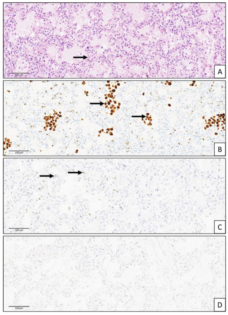
A less than one-month-old infant with symptoms of rhinitis died unexpectedly in his sleep. He was not born prematurely and had no known underlying disease. Cerebrospinal fluid, nasopharyngeal and lung samples, and rectal swab were found to be positive for subgroup A rhinovirus, while the blood was negative. This case highlights the important finding that the rhinovirus, a common pathogen associated with upper respiratory tract infections, can sometimes, as the only pathogen, lead to complications such as a cerebrospinal infection and be involved in the sudden infant death syndrome (SIDS). Vigilance is necessary in case of viral infections in the infant's environment, and measures of hygiene and protection must be encouraged in order to reduce the risk of the SIDS.
Keywords: SIDS; central nervous system; cerebrospinal fluid; fatal; infant; infection; rhinovirus; sudden death; virus.
Conflict of interest statement
The authors declare no conflicts of interest.
Figures
References
-
- van Benten I., Koopman L., Niesters B., Hop W., van Middelkoop B., de Waal L., van Drunen K., Osterhaus A., Neijens H., Fokkens W. Predominance of rhinovirus in the nose of symptomatic and asymptomatic infants. Pediatr. Allergy Immunol. 2003;14:363–370. doi: 10.1034/j.1399-3038.2003.00064.x. - DOI - PMC - PubMed
-
- Miller E.K., Williams J.V., Gebretsadik T., Carroll K.N., Dupont W.D., Mohamed Y.A., Morin L.L., Heil L., Minton P.A., Woodward K., et al. Host and viral factors associated with severity of human rhinovirus-associated infant respiratory tract illness. J. Allergy Clin. Immunol. 2011;127:883–891. doi: 10.1016/j.jaci.2010.11.041. - DOI - PMC - PubMed
Publication types
MeSH terms
LinkOut - more resources
Full Text Sources
Medical
