. 2024 Mar 31;16(4):549.
doi: 10.3390/v16040549.

A Bacterium-like Particle Vaccine Displaying Envelope Proteins of Canine Distemper Virus Can Induce Immune Responses in Mice and Dogs

Affiliations

A Bacterium-like Particle Vaccine Displaying Envelope Proteins of Canine Distemper Virus Can Induce Immune Responses in Mice and Dogs

Lina Liu et al. Viruses. .

Abstract

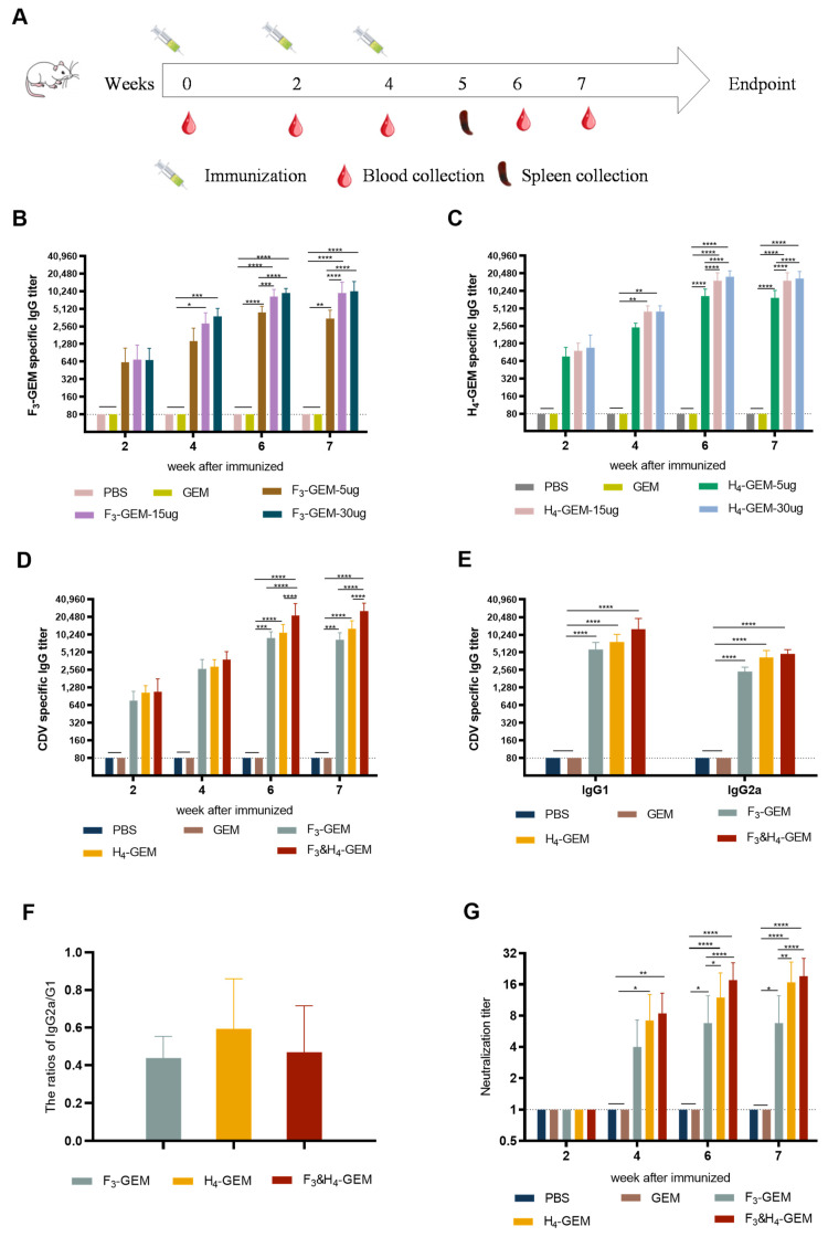
Canine distemper virus (CDV) can cause fatal infections in giant pandas. Vaccination is crucial to prevent CDV infection in giant pandas. In this study, two bacterium-like particle vaccines F3-GEM and H4-GEM displaying the trimeric F protein or tetrameric H protein of CDV were constructed based on the Gram-positive enhanced-matrix protein anchor (GEM-PA) surface display system. Electron microscopy and Western blot results revealed that the F or H protein was successfully anchored on the surface of GEM particles. Furthermore, one more bacterium-like particle vaccine F3 and H4-GEM was also designed, a mixture consisting of F3-GEM and H4-GEM at a ratio of 1:1. To evaluate the effect of the three vaccines, mice were immunized with F3-GEM, H4-GEM or F3 and H4-GEM. It was found that the level of IgG-specific antibodies and neutralizing antibodies in the F3 and H4-GEM group was higher than the other two groups. Additionally, F3 and H4-GEM also increased the secretion of Th1-related and Th2-related cytokines. Moreover, F3 and H4-GEM induce IgG and neutralizing antibodies' response in dogs. Conclusions: In summary, F3 and H4-GEM can provoke better immune responses to CDV in mice and dogs. The bacterium-like particle vaccine F3 and H4-GEM might be a potential vaccine candidate for giant pandas against CDV infection.

Keywords: CDV; F protein; H protein; bacterium-like particles; pandas; subunit vaccine.


Conflict of interest statement

The authors declare no conflicts of interest.

Figures

Figure 1
Figure 1
Construction and identification of F3-PA and H4-PA. (A) Schematic of the recombinant baculovirus expressing the F3-PA and H4-PA proteins. SP: signal peptide, T4: the trimeric motif; GCN4: the tetrameric motif; linker: (GGGGS)3; PA: indicates protein anchor. (B) IFA detection of F3-PA and H4-PA expression in baculovirus-infected Sf9 cells. Cells were infected with rBV-F3-PA or rBV-H4-PA. After 48 h, the cells were detected with a mouse anti-F or H protein polyclonal antibody. (C) Western blot analysis of F or H protein expression in Sf9-infected cells. M: molecular-weight marker, 1: cell sedimentation, 2: supernatant after sonication, and 3: uninfected cells. (D) Western blot identification of F3 and H4 proteins with non-reduction treatment; M: molecular-weight marker, 1: supernatant after sonication of F3-PA; 2: supernatant after sonication of H4-PA; (E) and analysis of the recombinant baculovirus titers. (E1): F3-PA; (E2): H4-PA; (E3,E4): Baculovirus control.
Figure 2
Figure 2
Identification of F3-GEM and H4-GEM. (A) TEM images of MG1363 Lactococcus lactis. (B) TEM images of GEM particles. (C) TEM images of F3-GEM. (D) TEM images of H4-GEM. (E) Western blot identification of F or H protein anchored on GEM particles. 1: F3-GEM; 2: H4-GEM. (F,G): 0.5 U GEM particles were mixed with 0 mL, 2 mL, 4 mL, 6 mL and 8 mL F or H protein. (H) The amount of F or H protein was determined. 1: GEM; 2: H4-GEM; 3: F3-GEMl; and 4–9: different concentrations of BSA proteins.
Figure 3
Figure 3
Serum antibody responses induced by F3-GEM and H4-GEM. Serum samples were collected at weeks 0, 2, 4, 6 and 7. Mouse specific total IgG, IgG1 and IgG2a antibody responses were measured using end-point dilution titers (n = 10 mice/group/time point). Data are shown as the mean ± SD and were analyzed by one-way ANOVA (* p < 0.05, ** p < 0.01, *** p < 0.001 and **** p < 0.0001). (A) Schematic of the experiment. (B,C) Analysis of serum specific total IgG titers induced by different doses of F3-GEM or H4-GEM. (D) The IgG titers induced via F3-GEM, H4-GEM or F3 and H4-GEM. (E): Analysis of IgG1 and IgG2a titers induced by the three vaccines. (F) The ratios of IgG2a/IgG1. (G) The neutralization antibody titers of the three vaccines.
Figure 4
Figure 4
ELISpot analysis of IFN-γ and IL-4 secretion via mouse splenocytes. The splenocytes were collected from each group 7 days after the third immunization was treated and analyzed. (A) The spots on the pictures showed the positive cells that secrete cytokines. The secretions of IFN-γ (B) and IL-4 (C) were measured by using an ELISpot kit. Data are shown as the mean ± SD and were analyzed using one-way ANOVA (**** p < 0.0001).
Figure 5
Figure 5
Detection of cytokines in splenocytes. Splenocytes were harvested from the mice 7 days after the third immunization and restimulated with F or H (10 µg/mL) in culture ex vivo. The concentrations of IFN-γ (A), IL-2 (B), TNF-α (C), IL-4 (D), IL-6 (E) and IL-10 (F) in the supernatant were measured with Meso Scale Discovery based on electrochemiluminescence. Data are shown as the mean ± SD and were analyzed using one-way ANOVA (*** p < 0.001 and **** p < 0.0001).
Figure 6
Figure 6
Antibodies in the immunized dogs. Data are shown as the mean ± SD and were analyzed by one-way ANOVA (* p < 0.05, *** p < 0.001, **** p < 0.0001). (A) The IgG-specific antibody titers in the serum of dogs. (B) The neutralization antibody titers in the serum of dogs.

Similar articles

Cited by

References

    1. Tao Y., Strelkov S.V., Mesyanzhinov V.V., Rossmann M.G. Rossmann, Structure of bacteriophage T4 fibritin: A segmented coiled coil and the role of the C-terminal domain. Structure. 1997;5:789–798. doi: 10.1016/S0969-2126(97)00233-5. - DOI - PubMed
    1. Yoshikawa Y., Ochikubo F., Matsubara Y., Tsuruoka H., Ishii M., Shirota K., Nomura Y., Sugiyama M., Yamanouchi K. Natural infection with canine distemper virus in a Japanese monkey (Macaca fuscata) Vet. Microbiol. 1989;20:193–205. doi: 10.1016/0378-1135(89)90043-6. - DOI - PubMed
    1. Appel M.J., Yates R.A., Foley G.L., Bernstein J.J., Santinelli S., Spelman L.H., Miller L.D., Arp L.H., Anderson M., Barr M., et al. Canine distemper epizootic in lions, tigers, and leopards in North America. J. Vet. Diagn. Investig. 1994;6:277–288. doi: 10.1177/104063879400600301. - DOI - PubMed
    1. Sheldon J.D., Cushing A.C., Wilkes R.P., Anis E., Dubovi E.J. Serologic response to canine distemper vaccination in captive linnaeus’s two-toed sloths (choloepus didactylus) after a fatal canine distemper virus outbreak. J. Zoo Wildl. Med. 2017;48:1250–1253. doi: 10.1638/1042-7260-48.4.1250. - DOI - PubMed
    1. Michelazzo M.M.Z., Oliveira T.E.S., Viana N.E., Moraes W., Cubas Z.S., Headley S.A. Immunohistochemical evidence of canine morbillivirus (canine distemper) infection in coatis (Nasua nasua) from Southern Brazil. Transbound. Emerg. Dis. 2020;67((Suppl. S2)):178–184. doi: 10.1111/tbed.13456. - DOI - PubMed

Publication types

MeSH terms