Chimeric Porcine Parvovirus VP2 Virus-like Particles with Epitopes of South African Serotype 2 Foot-and-Mouth Disease Virus Elicits Specific Humoral and Cellular Responses in Mice
- PMID: 38675963
- PMCID: PMC11054767
- DOI: 10.3390/v16040621
Chimeric Porcine Parvovirus VP2 Virus-like Particles with Epitopes of South African Serotype 2 Foot-and-Mouth Disease Virus Elicits Specific Humoral and Cellular Responses in Mice
Abstract
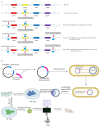
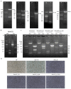
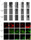
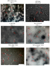
Southern Africa Territories 2 (SAT2) foot-and-mouth disease (FMD) has crossed long-standing regional boundaries in recent years and entered the Middle East. However, the existing vaccines offer poor cross-protection against the circulating strains in the field. Therefore, there is an urgent need for an alternative design approach for vaccines in anticipation of a pandemic of SAT2 Foot-and-mouth disease virus (FMDV). The porcine parvovirus (PPV) VP2 protein can embed exogenous epitopes into the four loops on its surface, assemble into virus-like particles (VLPs), and induce antibodies and cytokines to PPV and the exogenous epitope. In this study, chimeric porcine parvovirus VP2 VLPs (chimeric PPV-SAT2-VLPs) expressing the T-and/or B-cell epitopes of the structural protein VP1 of FMDV SAT2 were produced using the recombinant pFastBac™ Dual vector of baculoviruses in Sf9 and HF cells We used the Bac-to-Bac system to construct the recombinant baculoviruses. The VP2-VLP--SAT2 chimeras displayed chimeric T-cell epitope (amino acids 21-40 of VP1) and/or the B-cell epitope (amino acids 135-174) of SAT FMDV VP1 by substitution of the corresponding regions at the N terminus (amino acids 2-23) and/or loop 2 and/or loop 4 of the PPV VP2 protein, respectively. In mice, the chimeric PPV-SAT2-VLPs induced specific antibodies against PPV and the VP1 protein of SAT2 FMDV. The VP2-VLP-SAT2 chimeras induced specific antibodies to PPV and the VP1 protein specific epitopes of FMDV SAT2. In this study, as a proof-of-concept, successfully generated chimeric PPV-VP2 VLPs expressing epitopes of the structural protein VP1 of FMDV SAT2 that has a potential to prevent FMDV SAT2 and PPV infection in pigs.
Keywords: Epitope; FMDV; PPV; SAT2 serotype; chimeric VLP.
Conflict of interest statement
The authors declare no conflicts of interest.
Figures

Similar articles
-
Immunogenicity of adenovirus-derived porcine parvovirus-like particles displaying B and T cell epitopes of foot-and-mouth disease.Vaccine. 2016 Jan 20;34(4):578-585. doi: 10.1016/j.vaccine.2015.11.003. Epub 2015 Dec 9. Vaccine. 2016. PMID: 26685093
-
Potent neutralization activity against type O foot-and-mouth disease virus elicited by a conserved type O neutralizing epitope displayed on bovine parvovirus virus-like particles.J Gen Virol. 2019 Feb;100(2):187-198. doi: 10.1099/jgv.0.001194. Epub 2018 Dec 14. J Gen Virol. 2019. PMID: 30547855
-
Artificially designed hepatitis B virus core particles composed of multiple epitopes of type A and O foot-and-mouth disease virus as a bivalent vaccine candidate.J Med Virol. 2019 Dec;91(12):2142-2152. doi: 10.1002/jmv.25554. Epub 2019 Aug 24. J Med Virol. 2019. PMID: 31347713
-
B and T Cell Epitopes of the Incursionary Foot-and-Mouth Disease Virus Serotype SAT2 for Vaccine Development.Viruses. 2023 Mar 21;15(3):797. doi: 10.3390/v15030797. Viruses. 2023. PMID: 36992505 Free PMC article. Review.
-
Laboratory animal models to study foot-and-mouth disease: a review with emphasis on natural and vaccine-induced immunity.J Gen Virol. 2014 Nov;95(Pt 11):2329-2345. doi: 10.1099/vir.0.068270-0. Epub 2014 Jul 7. J Gen Virol. 2014. PMID: 25000962 Free PMC article. Review.
References
-
- WOAH. FAO Reference Laboratory Network for FMD. FMDV Prototype Strains. 2019. [(accessed on 14 May 2018)]. Available online: https://www.foot-and-mouth.org/FMDV-nomenclature-working-group/prototype....
Publication types
MeSH terms
Substances
Grants and funding
- 2021YFD1800300/the National Key R&D Program of China
- 32330107/the key project of the National Natural Science Foundation of China
- zujbky-2022-ct02/the Fundamental Research Funds of the Central Universities (Interdisciplinary Innovation Team Building Project)
- NCTIP-XD/C03/National Pig Technology Innovation Center
- 22ZD6NA012/the Development and Industrialization of Diagnosis of Important Livestock Diseases
- 22ZD6NA001/Basic Theoretical Research on Infection and Immune Prevention and Control of Important Livestock Diseases
- CAAS-CSLPDCP-2023002/Innovation Program of Chinese Academy of Agricultural Sciences
- CARS-35/the Construction of Agricultural Technology Industry System
- 2023SDZG02/the 14th Five-Year Plan Project of Guangdong Province
LinkOut - more resources
Full Text Sources
Research Materials
Miscellaneous

