. 2024 Apr 20;16(4):640.
doi: 10.3390/v16040640.

The Dual-Targeted Fusion Inhibitor Clofazimine Binds to the S2 Segment of the SARS-CoV-2 Spike Protein

Affiliations

The Dual-Targeted Fusion Inhibitor Clofazimine Binds to the S2 Segment of the SARS-CoV-2 Spike Protein

Matthew R Freidel et al. Viruses. .

Abstract

Clofazimine and Arbidol have both been reported to be effective in vitro SARS-CoV-2 fusion inhibitors. Both are promising drugs that have been repurposed for the treatment of COVID-19 and have been used in several previous and ongoing clinical trials. Small-molecule bindings to expressed constructs of the trimeric S2 segment of Spike and the full-length SARS-CoV-2 Spike protein were measured using a Surface Plasmon Resonance (SPR) binding assay. We demonstrate that Clofazimine, Toremifene, Arbidol and its derivatives bind to the S2 segment of the Spike protein. Clofazimine provided the most reliable and highest-quality SPR data for binding with S2 over the conditions explored. A molecular docking approach was used to identify the most favorable binding sites on the S2 segment in the prefusion conformation, highlighting two possible small-molecule binding sites for fusion inhibitors. Results related to molecular docking and modeling of the structure-activity relationship (SAR) of a newly reported series of Clofazimine derivatives support the proposed Clofazimine binding site on the S2 segment. When the proposed Clofazimine binding site is superimposed with other experimentally determined coronavirus structures in structure-sequence alignments, the changes in sequence and structure may rationalize the broad-spectrum antiviral activity of Clofazimine in closely related coronaviruses such as SARS-CoV, MERS, hCoV-229E, and hCoV-OC43.

Keywords: Arbidol; CHARMM; Clofazimine; Nsp13 helicase; S2 segment; S2 subunit; SARS-CoV-2; Spike-dependent; Toremifene; fusion inhibitor; molecular docking; surface plasmon resonance.


Conflict of interest statement

The authors declare no conflicts of interest.

Figures

Figure 1
Figure 1
Structure of fusion inhibitor derivatives used in this study.
Figure 2
Figure 2
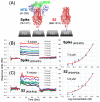
A direct SPR binding assay measures simultaneous binding to full-length Spike and the S2 segment. (A) Flow cell (FC) surfaces shown with attached Spike (FC2), reference FC1 and the S2 segment (FC4) and reference FC3. (B) Arbidol 1 binding to full-lennth Spike and (C) Arbidol 1 binding to the S2 segment showing both SPR sensorgrams and steady-state-affinity models.
Figure 3
Figure 3
Structure of the SARS-CoV-2 Spike S2 segment showing two possible fusion inhibitor binding sites. (A) The trimeric S2 segment in a pre-fusion conformation showing (B) Site 2 with Clofazimine 2 bound and (C) Site 1 with Arbidol 1 bound.
Figure 4
Figure 4
SPR data for fusion inhibitors binding to Spike and the S2 segment. Compounds from five different structural classes were shown to bind to the S2 segment. (A) Arbidol derivative 1c, (B) Arbidol derivative 1d, (C) Clofazimine 2, (D) Toremifene 3.
Figure 5
Figure 5
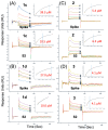
SPR duplicates for fusion inhibitors binding to the S2 segment. Duplicate binding curves for each compound are shown comparing only binding the S2 segment of Spike for (A) Arbidol 1, (B) Clofazimine 2, (C) Toremifene 3 and (D) Ecliptasaponin A 4.
Figure 6
Figure 6
Modeling fusion inhibitors at two possible binding sites on the S2 Segment. (A) Shown is the trimeric S2 segment in a prefusion conformation showing Site 2 with bound Clofazimine highlighted in purple and Site 1 with bound Arbidol highlighted in magenta. (B) For each fusion inhibitor, ∆Gbind values are calculated based on the lowest-energy cluster modeled at Site 1 and Site 2, shown in magenta and purple, respectively. Clofazimine 2 and Toremifene 3 are predicted to bind more favorably to Site 2, while Ecliptasaponin A 4, and OA Saponin 12a are predicted to bind more favorably to Site 1.
Figure 7
Figure 7
Predicted ∆Gbind values from Site 2 exhibit correlation with experimental SAR data for a series of Clofazimine derivatives. A series of 18 Clofazimine derivatives were well modeled as binding to Site 2, where sufficient linear correlation was achieved either comparing (A) all 18 derivatives (R2 = 0.264) or (B) two separate groups of compound series (R2 = 0.311) and (R2 = 0.306) with experimental SAR data. Poor correlation was observed when the compounds are modeled at Site 1 of S2, Nsp5, or Nsp16 “decoy” binding sites. When the series was modeled at the most favorable site on the Nsp13 helicase, the predicted ∆Gbind values exhibited some level of correlation for all 18 derivatives (R2 = 0.141) and quite reasonable correlation for the series of (15a, 15b, 15f, 15b, 15h) (R2 = 0.533).
Figure 8
Figure 8
Predicted ∆Gbind values from Site 2 correlated with experimental SAR data and explained SAR substitutions for all three R groups. A series of 18 Clofazimine derivatives were well modeled binding to Site 2 as two separate groups of compound series where the first series (A) shown in blue (6d, 6e, 7a, 7b, 7c, 7d, 7e, 7f, 7g, 7i, 7k, 7m, 7o) with correlation (R2 = 0.306) and the second series (B) shown in red (15a, 15b, 15f, 15b, 15h) had a slightly greater correlation (R2 = 0.311) with experimental SAR data. (C) A diagram to illustrate how the series modeled at Site 2 rationalized R group substitutions at R (purple), R1 (green) and R2 (cyan).
Figure 9
Figure 9
The predicted Site 2 is consistent with available structural information and may rationalize broad-spectrum activity of Clofazimine for MERS and other coronaviruses. (A) Predicted binding site for Clofazimine is shown illustrating the key binding site residues (W886, Q1036, K1038, V1040, and Y1047) shown below in the structure–sequence alignment. (B) A ribbon diagram is shown from a structure–sequence alignment between SARS-CoV-2 (6vxx.pdb) and MERS (8sak.pdb). (C) When docked at Site 2, the VFI tripeptide shows pharmacophore overlap with Clofazimine and the three major hydrophobic peptide side-chain pharmacophores. The surface model of the binding site is shown in (D) for SARS-CoV-2, showing a highly complementary binding surface for bound Clofazimine 2 in blue, where (E) shows that the binding surface shown in red is very similar in MERS with few atom clashes with the Clofazimine 2 binding mode. (F) Shows a sequence alignment derived from structure–sequence alignments with experimentally determined structures of the Spike protein from 15 different coronavirus strains. The sequence conservation of the SARS-CoV-2 residues that form the binding site (W886, A890, Q1036, K1038, V1040, Y1047 and R1107) are highlighted and denoted with the * symbol, where Q1036 is the most conserved of these residues.
Figure 10
Figure 10
A possible mechanism of action for binding at Site 2 is to stabilize the prefusion conformation and prevent conformational changes required for fusion. (A) Ribbon diagrams of the experimentally determined structures of the prefusion conformation and the superimposed post-fusion structure (6xra.pdb) of S2. The proposed binding site for Clofazimine 2 is highlighted with a magenta molecular surface. To visualize local conformational changes, four residue segments (943–1034), (1035–1070), (1078–1120) and (1121–1141) are shown as green, blue, cyan and red, respectively. (B) A zoomed-in molecular surface diagram showing the superimposed structure of the prefusion conformation in medium blue showing complementary molecular surface and interactions where, in the post-fusion structure, the magenta atoms and surface of 2 clash with the teal and yellow molecular surface that has undergone local hydrophobic collapse during the conformational transition.

References

    1. Wang X., Cao R., Zhang H., Liu J., Xu M., Hu H., Li Y., Zhao L., Li W., Sun X., et al. The anti-influenza virus drug, arbidol is an efficient inhibitor of SARS-CoV-2 in vitro. Cell Discov. 2020;6:28. doi: 10.1038/s41421-020-0169-8. - DOI - PMC - PubMed
    1. Xiao X., Wang C., Chang D., Wang Y., Dong X., Jiao T., Zhao Z., Ren L., Dela Cruz C.S., Sharma L., et al. Identification of Potent and Safe Antiviral Therapeutic Candidates Against SARS-CoV-2. Front. Immunol. 2020;11:586572. doi: 10.3389/fimmu.2020.586572. - DOI - PMC - PubMed
    1. Ke Y.Y., Peng T.T., Yeh T.K., Huang W.Z., Chang S.E., Wu S.H., Hung H.C., Hsu T.A., Lee S.J., Song J.S., et al. Artificial intelligence approach fighting COVID-19 with repurposing drugs. Biomed. J. 2020;43:355. doi: 10.1016/j.bj.2020.05.001. - DOI - PMC - PubMed
    1. Riva L., Yuan S., Yin X., Martin-Sancho L., Matsunaga N., Pache L., Burgstaller-Muehlbacher S., De Jesus P.D., Teriete P., Hull M.V., et al. Discovery of SARS-CoV-2 antiviral drugs through large-scale compound repurposing. Nature. 2020;586:113. doi: 10.1038/s41586-020-2577-1. - DOI - PMC - PubMed
    1. Yuan S., Yin X., Meng X., Chan J.F., Ye Z.W., Riva L., Pache L., Chan C.C., Lai P.M., Chan C.C., et al. Clofazimine broadly inhibits coronaviruses including SARS-CoV-2. Nature. 2021;593:418. doi: 10.1038/s41586-021-03431-4. - DOI - PubMed

Publication types

MeSH terms

LinkOut - more resources