. 2024 Jul 8;52(12):6811-6829.
doi: 10.1093/nar/gkae329.

CARM1 hypermethylates the NuRD chromatin remodeling complex to promote cell cycle gene expression and breast cancer development

Affiliations

CARM1 hypermethylates the NuRD chromatin remodeling complex to promote cell cycle gene expression and breast cancer development

Xue Chen et al. Nucleic Acids Res. .

Abstract

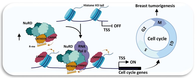
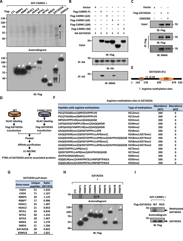
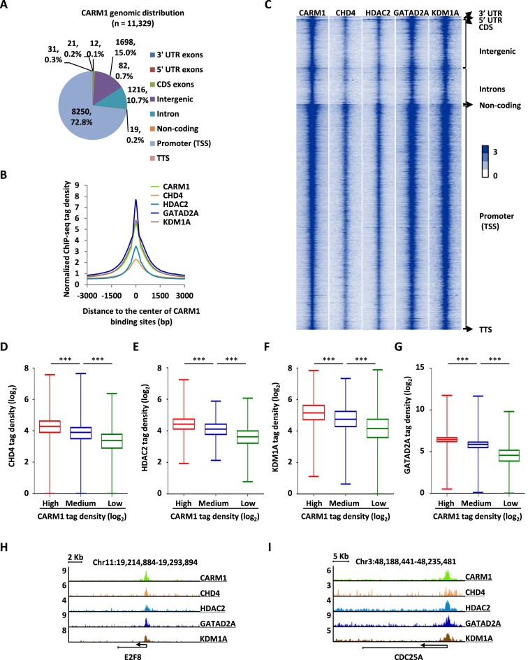
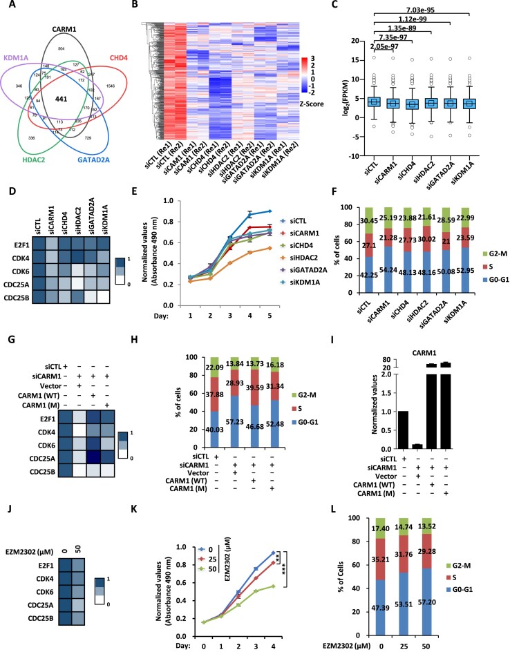
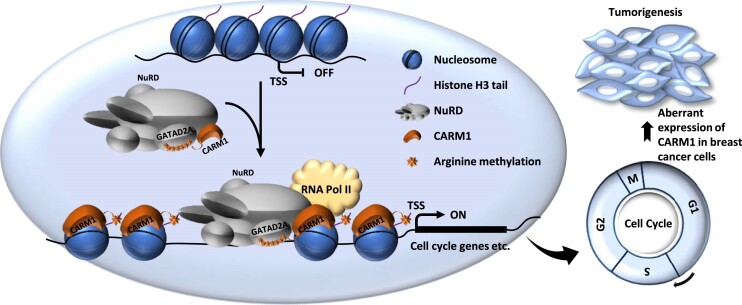
Protein arginine methyltransferase CARM1 has been shown to methylate a large number of non-histone proteins, and play important roles in gene transcriptional activation, cell cycle progress, and tumorigenesis. However, the critical substrates through which CARM1 exerts its functions remain to be fully characterized. Here, we reported that CARM1 directly interacts with the GATAD2A/2B subunit in the nucleosome remodeling and deacetylase (NuRD) complex, expanding the activities of NuRD to include protein arginine methylation. CARM1 and NuRD bind and activate a large cohort of genes with implications in cell cycle control to facilitate the G1 to S phase transition. This gene activation process requires CARM1 to hypermethylate GATAD2A/2B at a cluster of arginines, which is critical for the recruitment of the NuRD complex. The clinical significance of this gene activation mechanism is underscored by the high expression of CARM1 and NuRD in breast cancers, and the fact that knockdown CARM1 and NuRD inhibits cancer cell growth in vitro and tumorigenesis in vivo. Targeting CARM1-mediated GATAD2A/2B methylation with CARM1 specific inhibitors potently inhibit breast cancer cell growth in vitro and tumorigenesis in vivo. These findings reveal a gene activation program that requires arginine methylation established by CARM1 on a key chromatin remodeler, and targeting such methylation might represent a promising therapeutic avenue in the clinic.


Figures

Graphical Abstract
Graphical Abstract
Figure 1.
Figure 1.
CARM1 interacts with the NuRD complex. (A) HEK293 cells stably expressing Flag-HA-tagged CARM1 or empty vector were subjected to affinity purification, and the eluates were resolved by SDS-PAGE gel, silver-stained and subjected to mass spectrometry analysis. (B) Subunits in the NuRD complex and the corresponding number of unique peptides identified are shown as indicated. (C) HEK293 cells as described in (A) were subjected to immunoprecipitation (IP) with anti-Flag antibody followed by immunoblotting (IB) with antibodies as indicated. (D) In vitro methylation assay was performed by mixing core histones with eluates from control vector or Flag-tagged GATAD2A affinity purification in HEK293 cells, followed by autoradiogram (upper panel). Loading of core histones is shown by coomassie blue staining (CBS) (bottom panel). (E) GST pull-down assay was performed by mixing Flag-tagged GATAD2A purified from over-expressed HEK293 cells with GST or GST-tagged CARM1 followed by immunoblotting (IB) with anti-Flag antibody. (F, G) HEK293 cells were subjected to co-immunoprecipitation (Co-IP) assay with control IgG, anti-GATAD2A (F), or anti-CARM1 (G) antibody, followed by immunoblotting (IB) analysis with antibodies as indicated. (H) HEK293 cells transfected with control siRNA (siCTL) or siRNA specifically targeting GATAD2A (siGATAD2A) were subjected to immunoprecipitation (IP) with anti-CHD4 antibody, followed by immunoblotting (IB) with antibodies as indicated. (I) Schematic representation of full length (FL) and truncated CARM1 proteins. ΔN: amino (N)-terminus deletion; ΔC: carboxyl (C)-terminus deletion; C.C: catalytic core. (J) HEK293 cells were transfected with vectors expressing Flag and HA-tagged GATAD2A and empty vector, full length (FL), amino (N)-terminal deletion (ΔN), carboxyl (C)-terminal deletion (ΔC) or catalytic core only (C.C) CARM1 followed by immunoprecipitation (IP) with anti-HA antibody and immunoblotting (IB) with anti-Flag or anti-HA antibody. (K) HEK293 cells were transfected with vectors expressing HA-tagged GATAD2A and empty vector, full length (FL), or region 3-deleted (Δ3) CARM1 followed by immunoprecipitation (IP) with anti-HA antibody and immunoblotting (IB) with anti-Flag or anti-HA antibody.
Figure 2.
Figure 2.
CARM1 hypermethylates GATAD2A/2B in the NuRD complex. (A) In vitro methylation assay was performed by mixing GST-tagged CARM1 purified from bacterial cells with Flag-tagged, representative subunits in the NuRD complex including RBBP4, RBBP7, HDAC1, HDAC2, MTA1, MTA2 (truncation), MBD3, GATAD2A and KDM1A purified from over-expressed HEK293 cells, followed by autoradiogram to examine CARM1-mediated methylation (upper panel) or immunoblotting (IB) using anti-Flag antibody to examine the expression of the NuRD subunits (bottom panel). Black arrow and bracket (upper panel) indicated methylation of GATAD2A and its proteolytic fragments, respectively. White arrows indicated the size of the corresponding subunits in the NuRD complex. (B) In vitro methylation assay was performed by mixing purified GATAD2A with full length (FL), region 3-deleted (Δ3), region 4 deleted-(Δ4), amino (N)-terminal-deleted (ΔN), or enzymatic dead (M) CARM1, followed by immunoblotting (IB) with antibodies as indicated. (C) HEK293 cells were transfected with or without Flag-tagged GATAD2A and treated with or without EZM2302 (50 μM, 48 h), followed by immunoprecipitation (IP) with anti-Flag antibody and immunoblotting (IB) analysis with antibodies as indicated. (D) Schematic representation of the protocol applied for detecting differential binding proteins and post-translational modifications (PTMs) of GATAD2A in WT and CARM1 (KO) HEK293 cells. WT and CARM1 (KO) HEK293 cells were subjected to SILAC labeling and then transfected with vectors expressing Flag-tagged GATAD2A. Cells were then lysed, pooled and subjected to affinity purification using M2 agarose followed by mass spectrometry (MS) analysis. (E) Schematic representation of domain architecture of GATAD2A. Arginine methylation sites are shown by matchsticks. CR: Coil-coil region; FL: full-length. (F) Arginine methylation sites identified in GATAD2A through mass spectrometry analysis as shown in (C). me1: mono-methylation; me2: di-methylation. (G) List of subunits in the CARM1 and NuRD complex identified to be associated with GATAD2A by mass spectrometry (MS) analysis, and the number of peptides and the ratio of the abundance of each subunit in WT and CARM1 (KO) cells are shown. (H) In vitro methylation assay was performed by mixing Flag-tagged GATAD2A with PRMT proteins purified from over-expressed HEK293 cells as indicated, followed by autoradiogram (upper panel) or immunoblotting (IB) using anti-Flag antibody (bottom panel). White arrows indicated the expression of all PRMTs. Star indicated methylation of GATAD2A. (I) In vitro methylation assay was performed by mixing GST-tagged CARM1 purified from bacterial cells with Flag-tagged wild-type (WT) or GATAD2A mutant with seven arginine methylation sites substituted with lysines (7R/K) purified from over-expressed HEK293 cells, followed by autoradiogram (upper panel) or immunoblotting (IB) using anti-Flag antibody (bottom panel).
Figure 3.
Figure 3.
CARM1 and NuRD complex occupy a large number of chromatin sites in common. (A) Genomic distribution of CARM1 binding sites identified by ChIP-seq. (B, C) Histogram (B) and heat map (C) representation of CARM1, CHD4, HDAC2, GATAD2A and KDM1A ChIP-seq tag density centered on CARM1 binding sites. bp: base pair. (D–G) Box plot representation of ChIP tag density (log2) of CHD4 (D), HDAC2 (E), KDM1A (F) and GATAD2A (G) on CARM1 binding sites, which were divided into three sub-classes, high, medium and low based on ChIP-seq tag density (± s.e.m., ***P < 0.001). (H, I) Genome browser views of CARM1, CHD4, HDAC2, GATAD2A and KDM1A ChIP-seq on E2F8 (H) and CDC25A (I) genes.
Figure 4.
Figure 4.
CARM1 and NuRD activate a large set of cell cycle genes to promote cell cycle progression in a CARM1 enzymatic activity-dependent manner. (A) HEK293 cells were transfected with control siRNA (siCTL) or siRNA specific against CARM1 (siCARM1) or representative subunits in NuRD including CHD4, HDAC2, GATAD2A and KDM1A (siCHD4, siHDAC2, siGATAD2A and siKDM1A) followed by RNA-seq analysis. Genes positively-regulated by CARM1, CHD4, HDAC2, GATAD2A, and KDM1A in common are shown by venn diagram (q < 0.05). (B, C) The expression levels (FPKM, log2) for those 441 genes as described in (A) are shown by heat map (B) and box plot (C). (D) HEK293 cells as described in (A) were subjected to RT-qPCR analysis to examine the expression of selected cell cycle genes as indicated, and data was represented by heat map. (E, F) HEK293 cells as described in (A) were subjected to cell proliferation assay (E) and FACS analysis (F). (G–I) HEK293 cells were transfected with control siRNA (siCTL) or siRNA specifically targeting CARM1 (siCARM1) in the presence or absence of control vector or vector expressing wild-type CARM1 (WT) or its enzymatically deficient mutant (M), followed by RT-qPCR analysis to examine the expression of selected cell cycle genes as indicated (G) and CARM1 (I), and FACS analysis to check cell cycle progression (H). (J) HEK293 cells treated with or without EZM2302 (50 μM, 48 h) were subjected to RNA extraction and RT-qPCR analysis to examine the expression of selected cell cycle genes as indicated. (K, L) HEK293 cells treated with or without EZM2302 at concentration as indicated were subjected to cell proliferation assay (K) and FACS analysis (L) (± s.e.m., **P < 0.01, ***P < 0.001).
Figure 5.
Figure 5.
CARM1-mediated hypermethylation of GATAD2A is involved in NuRD chromatin binding, transcriptional activation of cell cycle genes, and cell cycle progression. (A, B) GATAD2A ChIP-seq was performed in WT or CARM1 (KO) HEK293 cells, and heat map (A) and box plot (B) representation of GATAD2A ChIP-seq tag density centered on transcription start sites (TSSs) of genes positively-regulated by CARM1 and NuRD is shown. (C, D) Genome browser views of CARM1 and GATAD2A ChIP-seq, either in WT or CARM1 (KO) HEK293 cells, on E2F8 (C) and CDC25A (D) gene is shown. (E) WT or CARM1 (KO) HEK293 cells were subjected to ChIP with anti-IgG or anti-GATAD2A specific antibody followed by qPCR analysis with primers specifically targeting promoter regions of selected cell cycle genes as indicated (± s.e.m., *P < 0.05, ***P < 0.001). (F, G) HEK293 cells were transfected with siRNA targeting 3′UTR region of GATAD2A together with or without control vector or vectors expressing HA-tagged WT or mutant GATAD2A (7R/K), followed by ChIP-seq with anti-HA antibody. Heat map (F) and box plot (G) representation of ChIP-seq tag density of WT or mutant GATAD2A (7R/K) HEK293 cells centered on transcription start sites (TSSs) of genes positively-regulated by CARM1 and NuRD. (H, I) Genome browser views of GATAD2A WT and GATAD2A mutant (7R/K) ChIP-seq on E2F8 (H) and CDC25A (I) gene are shown. (J) HEK293 cells as described in (F) were subjected to ChIP-qPCR analysis with primers specifically targeting the promoter region of selected cell cycle genes as indicated (± s.e.m., **P < 0.01, ***P < 0.001). (K) HEK293 cells were transfected with control siRNA or siRNA targeting 3′UTR region of GATAD2A together with or without control vector or vectors expressing HA-tagged WT or mutant GATAD2A (7R/K), followed by RT-qPCR analysis to examine the expression of selected cell cycle genes as indicated, and data was represented by heat map. (L) HEK293 cells as described in (K) were subjected to FACS analysis. (M) HEK293 cells as described in (K) were subjected to RT-qPCR analysis to examine the expression of GATAD2A.
Figure 6.
Figure 6.
CARM1-mediated GATAD2A methylation is required for breast cancer cell growth both in vitro and in vivo. (A, B) MCF7 cells were subjected to co-immunoprecipitation (Co-IP) assay with control IgG, anti-GATAD2A (A), or anti-CARM1 (B) antibody and followed by immunoblotting (IB) with antibodies as indicated. (C) MCF7 cells were transfected with siRNA (siCTL) or siRNA specific against CARM1 (siCARM1) and then infected with lenti-viral vector expressing Flag-tagged GATAD2A, followed by immunoprecipitation (IP) with anti-Flag antibody and immunoblotting (IB) analysis with antibodies as indicated. (D) MCF7 cells transfected with siRNA (siCTL) or siRNA specific against CARM1 (siCARM1) were subjected to ChIP with control IgG or anti-GATAD2A specific antibody, followed by qPCR analysis with primers specifically targeting promoter regions of selected cell cycle genes as indicated (± s.e.m., ***P < 0.001). (E-H) MCF7 cells transfected with control siRNA (siCTL) or siRNA specific against CARM1 (siCARM1) or GATAD2A (siGATAD2A) were subjected to RNA extraction and RT-qPCR analysis to examine the expression of selected cell cycle genes as indicated (E), cell proliferation assay (F), FACS analysis (G), and colony formation assay (H) (± s.e.m., ***P < 0.001). (I) Quantification of the crystal violet dye as shown in (H). (J) MCF7 cells were infected with control shRNA (shCTL) or shRNA targeting 3′UTR region of GATAD2A together with or without control lentiviral vector or vectors expressing WT or mutant GATAD2A (7R/K), followed by cell proliferation assay (± s.e.m., ***P < 0.001). (K) MCF7 cells infected with control shRNA (shCTL) or shRNA specific against CARM1 or GATAD2A, were injected subcutaneously into female BALB/C nude mice and brushed with estrogen (E2, 10−2 M) on the neck every two days for six weeks. Mice were then euthanized and tumors were excised and weighted (± s.e.m., *P < 0.05, ***P < 0.001). (L) MCF7 cells as described in (E) were injected subcutaneously into female BALB/C nude mice and brushed with estrogen (E2, 10−2 M) on the neck every two days for six weeks. Mice were then euthanized and tumors were excised and weighted (± s.e.m., *P < 0.05, N.S., not significant).
Figure 7.
Figure 7.
Targeting CARM1 with EZM2302 inhibits breast cancer cell growth both in vitro and in vivo. (A) MCF7 cells were transfected with or without Flag-tagged GATAD2A and treated with or without EZM2302 (25 μM, 48 h), followed by immunoprecipitation (IP) with anti-Flag antibody and immunoblotting (IB) analysis with antibodies as indicated. (B) MCF7 cells treated with or without EZM2302 (25 μM, 48 h) were subjected to ChIP with control IgG or anti-GATAD2A specific antibody followed by qPCR analysis with primers specifically targeting promoter regions of selected cell cycle genes as indicated (± s.e.m., *P < 0.05, ***P < 0.001). (C) MCF7 cells treated with or without EZM2302 (25 μM, 48 h) were subjected to RNA extraction and RT-qPCR analysis to examine the expression of selected cell cycle genes as indicated (± s.e.m., *P < 0.05, **P < 0.01, ***P < 0.001). (D-F) MCF7 cells treated with or without EZM2302 at concentration as indicated were subjected to cell proliferation assay (D), FACS analysis (E), and colony formation assay (F) (± s.e.m., **P < 0.01, ***P < 0.001). (G) Quantification of the crystal violet dye as shown in (F) (± s.e.m., ***P < 0.001). (H) MCF7 cells were injected subcutaneously into female BALB/C nude mice, and brushed with estrogen (E2, 10−2 M) on the neck every two days until tumor size reached approximately 100 mm3. Mice were then randomly assigned into three groups and treated with or without EZM2302 intraperitoneally every two days for 13 days. Tumors were harvested, photographed, and weighted. (I) The weight of tumors in (H) is shown (± s.e.m., **P < 0.01, ***P < 0.001). (J) The growth curve of tumors in (H) is shown. (K) Tumors as described in (H) were subjected to RNA extraction and RT-qPCR analysis to examine the expression of selected cell cycle genes as indicated (± s.e.m., *P < 0.05, **P < 0.01, ***P < 0.001).
Figure 8.
Figure 8.
A proposed model of CARM1-mediated NuRD methylation in gene transcriptional regulation and cell cycle control. CARM1 and NuRD commonly bind and activate a large cohort of genes with implications in cell cycle control to facilitate G1/S transition. Activation of this gene program requires CARM1 to methylate a key subunit in NuRD, GATAD2A/2B. Aberrant expression of CARM1 and NuRD results in uncontrolled expression of these cell cycle genes and cell cycle progression, leading to cancers, such as breast cancer.

Similar articles

Cited by

References

    1. Chen D., Ma H., Hong H., Koh S.S., Huang S.M., Schurter B.T., Aswad D.W., Stallcup M.R.. Regulation of transcription by a protein methyltransferase. Science. 1999; 284:2174–2177. - PubMed
    1. Teyssier C., Chen D., Stallcup M.R.. Requirement for multiple domains of the protein arginine methyltransferase CARM1 in its transcriptional coactivator function. J. Biol. Chem. 2002; 277:46066–46072. - PubMed
    1. Larsen S.C., Sylvestersen K.B., Mund A., Lyon D., Mullari M., Madsen M.V., Daniel J.A., Jensen L.J., Nielsen M.L.. Proteome-wide analysis of arginine monomethylation reveals widespread occurrence in human cells. Sci. Signal. 2016; 9:rs9. - PubMed
    1. Shishkova E., Zeng H., Liu F., Kwiecien N.W., Hebert A.S., Coon J.J., Xu W.. Global mapping of CARM1 substrates defines enzyme specificity and substrate recognition. Nat. Commun. 2017; 8:15571. - PMC - PubMed
    1. Peng B.L., Li W.J., Ding J.C., He Y.H., Ran T., Xie B.L., Wang Z.R., Shen H.F., Xiao R.Q., Gao W.W.et al. .. A hypermethylation strategy utilized by enhancer-bound CARM1 to promote estrogen receptor alpha-dependent transcriptional activation and breast carcinogenesis. Theranostics. 2020; 10:3451–3473. - PMC - PubMed

MeSH terms

Substances