. 2024 Jun:175:116666.
doi: 10.1016/j.biopha.2024.116666. Epub 2024 Apr 27.

The e-liquid flavoring cinnamaldehyde induces cellular stress responses in human proximal tubule (HK-2) kidney cells

Affiliations

The e-liquid flavoring cinnamaldehyde induces cellular stress responses in human proximal tubule (HK-2) kidney cells

Ashley Cox et al. Biomed Pharmacother. 2024 Jun.

Abstract

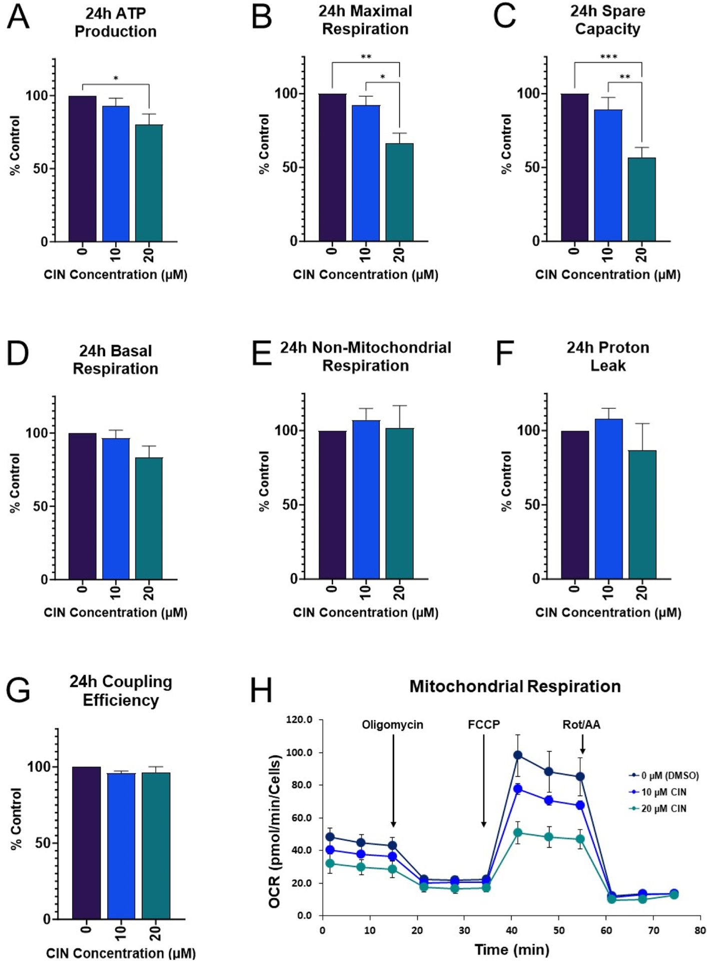
Flavored e-liquid use has become popular among e-cigarette users recently, but the effects of such products outside the lung are not well characterized. In this work, acute exposure to the popular flavoring cinnamaldehyde (CIN) was performed on human proximal tubule (HK-2) kidney cells. Cells were exposed to 0-100 µM CIN for 24-48 h and cellular stress responses were assessed. Mitochondrial viability via MTT assay was significantly decreased at 20 µM for 24 and 48 h exposure. Seahorse XFp analysis showed significantly decreased mitochondrial energy output at 20 µM by 24 h exposure, in addition to significantly reduced ATP Synthase expression. Seahorse analysis also revealed significantly decreased glycolytic function at 20 µM by 24 h exposure, suggesting inability of glycolytic processes to compensate for reduced mitochondrial energy output. Cleaved caspase-3 expression, a mediator of apoptosis, was significantly increased at the 24 h mark. C/EBP homologous protein (CHOP) expression, a mediator of ER-induced apoptosis, was induced by 48 h and subsequently lost at the highest concentration of 100 µM. This decrease was accompanied by a simultaneous decrease in its downstream target cleaved caspase-3 at the 48 h mark. The autophagy marker microtubule-associated protein 1 A/1B light chain 3 (LC3B-I and LC3B-II) expression was significantly increased at 100 µM by 24 h. Autophagy-related 7 (ATG7) protein and mitophagy-related proteins PTEN-induced putative kinase 1 (PINK1) and PARKIN expression were significantly reduced at 24 and 48 h exposure. These results indicate acute exposure to CIN in the kidney HK-2 model induces mitochondrial dysfunction and cellular stress responses.

Keywords: Cellular stress; Cinnamaldehyde; E-cigarette; Flavor; Kidney.


Conflict of interest statement

Declaration of Competing Interest The authors declare that they have no known competing financial interests or personal relationships that could have appeared to influence the work reported in this paper

Figures

Fig. 1.
Fig. 1.. Cinnamaldehyde chemical structure.
Cinnamaldehyde (CAS 104–55–2) is an α,β-unsaturated aldehyde used as a flavoring agent.
Fig. 2.
Fig. 2.. Cinnamaldehyde effects on cell and mitochondrial viability.
CIN diminished significantly mitochondrial activity at 24 (A) and 48 h (B) (p < 000.1) starting at 20 μM. Statistical difference from 0 μM (DMSO) vehicle control group is indicated by different superscript letters (a-g). Trypan blue exclusion showed no significant differences between control and treatment groups (C, D). One-way ANOVA was used for statistical analysis followed by a Tukey post-hoc test. Values represent mean ± SEM for a minimum of 4 independent experiments using different biological replicates.
Fig. 3.
Fig. 3.. Cinnamaldehyde effects on mitochondrial function.
At 24 h, 20 μM CIN significantly decreased ATP production, maximal respiration, and spare respiratory capacity when compared to control (A-C). No significant differences in basal respiration, non-mitochondrial respiration, proton leak, or coupling efficiency were observed (D-G). Representative time course profile of a Seahorse cell mito stress test following exposure to CIN for 24 h (H). Total number of cells was measured using the CyQUANT Direct Cell Proliferation Assay and results were normalized to number of cells. Statistical differences denoted by an asterisk (* p < 0.05, ** p < 0.01, *** p < 0.001). Values represent mean ± SEM for a minimum of 4 independent experiments using different biological replicates.
Fig. 4.
Fig. 4.. Cinnamaldehyde effects on glycolytic function.
After 24 h, 20 μM CIN significantly decreased glycolysis, glycolytic capacity, and glycolytic reserve when compared to control (A-C). Non-glycolytic acidification was decreased, but not significantly (D). Representative time course profile of a Seahorse cell glycolysis stress test following exposure to CIN for 24 h (H). Total number of cells was measured using the CyQUANT Direct Cell Proliferation Assay and results were normalized to number of cells. Statistical differences denoted by an asterisk (** p < 0.01, *** p < 0.001, **** p < 0.0001). Values represent mean ± SEM for a minimum of 4 independent experiments using different biological replicates.
Fig. 5.
Fig. 5.. Cinnamaldehyde effects on mitochondrial OXPHOS complexes.
CIN exposure significantly reduced complex V expression at 24 and 48 h (A-B). Expression of complexes I and II were significantly reduced only between non-control groups and 100 μM (C-D). Results were normalized to total protein concentration. The positive control was rat heart lysate. Statistical differences denoted by an asterisk (* p < 0.05, ** p < 0.01). One-way ANOVA was used for statistical analysis followed by a Tukey post-hoc test. Values represent mean ± SEM for a minimum of 4 independent experiments using different biological replicates.
Fig. 6.
Fig. 6.. Cinnamaldehyde effects on ER stress and apoptotic pathway.
CHOP expression was triggered by 48 h exposure to CIN, and significantly decreased at 100 μM (A). Comparison of CHOP expression at 24 h vs. 48 h (B). Cleaved caspase-3 significantly increased after 24 h exposure to CIN (C), however this expression was lost by 48 h (D). Results were normalized to total protein concentration. The +CON for CHOP was C2C12 cells treated with thapsigargin. The +/−CON for cleave caspase 3 were HeLa cells treated with or without staurosporine. Statistical differences denoted by an asterisk (* p < 0.05, ** p < 0.01, *** p < 0.001). One-way ANOVA was used for statistical analysis followed by a Tukey post-hoc test. Values represent mean ± SEM for a minimum of 4 independent experiments using different biological replicates.
Fig. 7.
Fig. 7.. Cinnamaldehyde effects on autophagy marker LC3B.
CIN significantly increased LC3B-I and LC3B-II expression at 24 and 48 h (A-B, D-E). The ratio of LC3B II to LC3B-I was significantly increased at 24 h, and an increasing but non-significant trend was observed at 48 h (C, F). Comparison of LC3B-I and LC3B-II expression at 24 and 48 h (G). Results were normalized to total protein concentration. The −/+CON were HeLa cells treated with DMSO or staurosporine. Statistical differences denoted by an asterisk (* p < 0.05, ** p < 0.01, *** p < 0.001, **** p < 0.0001). One-way ANOVA was used for statistical analysis followed by a Tukey post-hoc test. Values represent mean ± SEM for a minimum of 4 independent experiments using different biological replicates.
Fig. 8.
Fig. 8.. Cinnamaldehyde effects on autophagy-related 7 protein.
After 24 h exposure to CIN, expression of ATG7 significantly decreased between non-control groups and 100 μM CIN (A). ATG7 expression significantly decreased at 48 h between control and 100 μM (B). Results were normalized to total protein concentration. HeLa cell lysate from cells treated with staurosporine was used as the positive control. Statistical differences denoted by an asterisk (* p < 0.05, ** p < 0.01). One-way ANOVA was used for statistical analysis followed by a Tukey post-hoc test. Values represent mean ± SEM for a minimum of 4 independent experiments using different biological replicates.
Fig. 9.
Fig. 9.. Cinnamaldehyde effects on mitophagy-related proteins PINK1 and PARKIN.
Both 24 and 48 h exposure significantly reduced expression of PINK1 and PARKIN (A-D). PINK1 cleavage (A, bottom) at 24 h is decreased by 48 h mark (C, bottom). Results were normalized to total protein concentration. PINK1 positive control was HeLa cell lysate treated with staurosporine while mouse brain lysate was used as the positive control for Parkin Statistical differences denoted by an asterisk (* p < 0.05). One-way ANOVA was used for statistical analysis followed by a Tukey post-hoc test. Values represent mean ± SEM for a minimum of 4 independent experiments using different biological replicates.

Similar articles

Cited by

References

    1. Birdsey J, Cornelius M, Jamal A, et al., Tobacco Product Use Among U.S. Middle and High School Students - National Youth Tobacco Survey, 2023, MMWR Morb. Mortal. Wkly Rep 72 (2023) 1173–1182, 10.15585/mmwr.mm7244a1. - DOI - PMC - PubMed
    1. Administration, Fa.D., FDA finalizes enforcement policy on unauthorized flavored cartridge-based e-cigarettes that appeal to children including fruit and mint.pdf. 2020.
    1. Miech RA, Johnston LD, Patrick ME, O’Malley PM, Bachman JG, Schulenberg JE, Monitoring the Future national survey results on drug use, 1975–2022: Secondary school students. Monitoring the Future Monograph Series. Ann Arbor, MI: Institute for Social Research, University of Michigan. Available at https://monitoringthefuture.org/results/publications/monographs/2023.
    1. Chaffee BW, Couch ET, Wilkinson ML, Donaldson CD, Cheng NF, Ameli N, Zhang X, Gansky SA, Flavors increase adolescents’ willingness to try nicotine and cannabis vape products, Drug Alcohol Depend. 246 (2023) 109834, 10.1016/j.drugalcdep.2023.109834. - DOI - PMC - PubMed
    1. Pepper JK, Ribisl KM, Brewer NT, Adolescents’ interest in trying flavoured e-cigarettes, Tob. Control 25 (Suppl 2) (2016) ii62–ii66, 10.1136/tobaccocontrol-2016-053174. - DOI - PMC - PubMed

MeSH terms