. 2024 May 14;57(5):1071-1086.e7.
doi: 10.1016/j.immuni.2024.04.003. Epub 2024 Apr 26.

CD80 on skin stem cells promotes local expansion of regulatory T cells upon injury to orchestrate repair within an inflammatory environment

Affiliations

CD80 on skin stem cells promotes local expansion of regulatory T cells upon injury to orchestrate repair within an inflammatory environment

Jingyun Luan et al. Immunity. .

Abstract

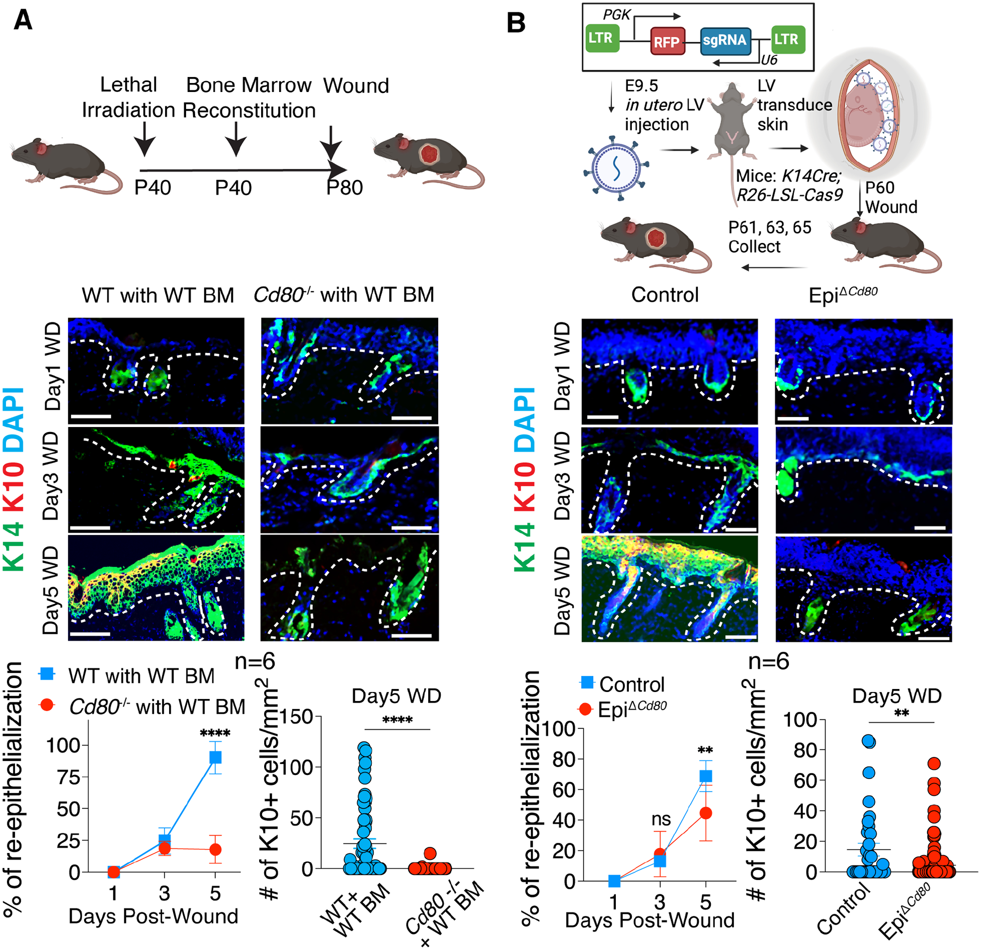
Following tissue damage, epithelial stem cells (SCs) are mobilized to enter the wound, where they confront harsh inflammatory environments that can impede their ability to repair the injury. Here, we investigated the mechanisms that protect skin SCs within this inflammatory environment. Characterization of gene expression profiles of hair follicle SCs (HFSCs) that migrated into the wound site revealed activation of an immune-modulatory program, including expression of CD80, major histocompatibility complex class II (MHCII), and CXC motif chemokine ligand 5 (CXCL5). Deletion of CD80 in HFSCs impaired re-epithelialization, reduced accumulation of peripherally generated Treg (pTreg) cells, and increased infiltration of neutrophils in wounded skin. Importantly, similar wound healing defects were also observed in mice lacking pTreg cells. Our findings suggest that upon skin injury, HFSCs establish a temporary protective network by promoting local expansion of Treg cells, thereby enabling re-epithelialization while still kindling inflammation outside this niche until the barrier is restored.

Keywords: CD80; Treg cell; hair follicle stem cell; neutrophils; wound repair.


Conflict of interest statement

Declaration of interests E.F. is on the editorial board of some Elsevier Journals (Cell, Cell Stem Cell, and Developmental Cell). A.Y.R. is a member of SAB and has equity in Coherus, RAPT Therapeutics, Sonoma Biotherapeutics, Santa Ana Bio, and Vedanta Biosciences; is an SAB member of BioInvent and Amgen; and holds a therapeutic Treg cell depletion IP licensed to Takeda.

Figures

Figure 1:
Figure 1:. Skin epithelium activates an immune-modulatory program during wound healing.
A) Schematic showing the lineage tracing of basal skin epithelium, followed by partial thickness wounding. B) Gene ontology analysis of differentially expressed genes in basal skin keratinocytes from wounded skin (day3 wound) compared to cells from unwounded skin. C) Heat map and representative genes that are differentially expressed in basal skin keratinocytes from wounded skin (day3 wound) compared to cells from unwounded skin. D) UMAP plots showing expression of various markers, including Cd80 and MHCII genes, activated in the basal epithelial skin cells during wound repair. E) Flow cytometry plots and MFI quantification of CD80 and MHCII on various cell populations in unwounded (UD) or wounded (WD) skin. Each symbol represents an independent animal. Representative data from three independent experiments are shown. Unpaired t test, * p<0.05; ** p<0.01. See also Figure S1. F) ImageStream analysis to visualize co-expression of CD80 and MHCII in the Tomato+ basal epithelial stem cells that have acquired Integrin α5+ and migrated into the wounded skin (day3 wound).
Figure 2:
Figure 2:. CD80 is activated in a subset of HFSCs that enter the wound bed during wound repair.
A) IF images of sagittal sections of partial thickness wounds showing the healing dynamics of Sox9+ HFSCs that were lineage-traced in Sox9-CreER; Rosa26-tdTomato mice. Scale bars: 100 μm B) IF images of sagittal sections of unwounded or wounded (day 3) skin showing that Tomato+ HFSC cells that acquire a5 are also positive for CD80. Scale bars: 100 μm C) Flow cytometry analysis and MFI quantification of CD80 and MHCII on on various cell populations in unwounded (UD) or wounded (WD) skin. Each symbol represents an independent animal. Representative data from three independent experiments are shown. Unpaired t test, ** p<0.01. D) ImageStream analysis to visualize CD80 and MHCII on migratory Tomato+α5+ HFSCs from wounded skin (day3 wound) on Sox9CreER traced mice. E) ATAC-sequencing peaks showing the accessibility status of the Cd80 and MHCII (H2-Aa and H2-Ab1) genomic loci within the chromatin of FACS-purified, Sox9CreER-lineage traced HFSCs from unwounded or wounded skin (day3 wound).
Figure 3:
Figure 3:. Specific ablation of CD80 in HFSCs results in wound healing delays.
A) Schematic is of wounding in irradiated Cd80−/− or WT control mice and reconstituted with WT bone marrow (BM). Shown below are IF images and quantifications of the percentage of wound re-epithelialization (K14+) and the number of differentiated suprabasal cells (K10+) in the skins at times indicated after partial thickness wounding. B) Schematic is of lentivirus injected into the amniotic sac of E9.5 embryos of Krt14-Cre; Rosa26-LSL-Cas9 mice to specifically transduce surface epithelial skin progenitors, here with Cd80-targeting sgRNAs (EpiΔCd80) or use Cre negative mice as controls. Shown below are IF images and quantifications of the percentage of wound re-epithelialization (K14+) and the number of differentiated suprabasal cells (K10+) in the skins at times indicated after partial thickness wounding. Each symbol represents a technical replicate. Results pooled from two independent experiments are shown (n=3 animals for each time point in each experiment). Unpaired t test, ** p<0.01; **** p<0.0001. Scale bars: 100 μm. See also Figure S2.
Figure 4:
Figure 4:. Wound-induced migrating stem cells express CD80 to stimulate local Treg expansion and create a protective niche around the re-epithelializing tissue.
(A to C) Total CD4 T cells (A), Foxp3+ Treg cells (B) and Foxp3- Tconv cells (C) are quantified by flow cytometry in unwounded (UD) or wounded (WD) skin at day5 following injury. n=9 pooled from three independent experiments. Each symbol represents an individual animal. Pooled quantifications from three independent experiments are shown. D) Schematic and flow cytometry quantification below shows the efficient Treg depletion. IF and quantification shows re-epithelialization (K14+) and differentiation (K10+) in mice with or without Treg depletion. E) IF images of total GFP+CD3+ Tregs (quantified at right) that infiltrate the skin wounds of WT or Cd80−/− mice reconstituted with bone marrow from Foxp3-GFP Treg reporter mice. Each symbol represents a technical replicate. Representative images and pooled quantifications from two independent experiments are shown. F) Quantification of the distance between GFP+ Treg cells that enter/expand within the wound bed in (E), from the migrating epithelial stem cells. Each symbol represents a technical replicate. Unpaired t test, ** p<0.01; *** p<0.001. Scale bars: 100 μm. See also Figure S3.
Figure 5:
Figure 5:. CD80 is important for skin stem cells to suppress neutrophil infiltration into the re-epithelializing wound bed.
A) Hematoxylin and eosin staining of the wounded skin from WT or Cd80−/− mice reconstituted with WT bone marrow. Representative images from two independent experiments are shown. Scale bar: 100 μm. B) Ultrastructural images of the wounded skin from WT or Cd80−/− mice reconstituted with WT bone marrow. Arrows point to polymorphonuclear cells. Neutrophils are color-coded in green. The re-epithelialized skin is color-coded in yellow. Representative images from two independent experiments are shown. Scale bar: 10 μm. C) Flow cytometry quantification of the number of Ly6GHi neutrophils within the CD11b+MHCIILow immune population in Day5 wounds on control or EpiΔCd80 mice. Each symbol represents an individual animal. Pooled quantifications from three independent experiments are shown. D) IF images and quantifications of Ly6G+ neutrophils per mm2 in wounded skin from WT or Cd80−/− mice reconstituted with WT bone marrow. Representative images and pooled quantifications from two independent experiments are shown. Scale bar: 100 μm. Unpaired t test, * p<0.05; ** p<0.01.
Figure 6.
Figure 6.. CD80 expression by migrating epithelial stem cells promotes the expansion of extrathymically-induced Treg cells during wound healing.
A) Schematic of experimental design and flow cytometry quantification of the frequency and number of preexisting (double positive: Tomato+GFP+) and newly induced (single positive: GFP+Tomato) Treg cells in day 5 wounds (n=3 in each experiment). Representative flow cytometry plots and pooled quantifications from two independent experiments are shown. B to D) IF images and quantification of re-epithelization (B), flow cytometry quantifications of the Treg cells (C) and neutrophils (D) in the Day5 wounds from skins of Rag2−/− mice that were reconstituted with Foxp3GFP control or Foxp3ΔCNS1-GFP bone marrow. Scale bar: 100 μm. Representative images and pooled quantifications from two independent experiments are shown. E to G) Image quantification of re-epithelization (E) and flow cytometry quantifications of the Treg cells (F) and neutrophils (G) in the Day5 wounds from skins of WT or Cd80−/− mice that were reconstituted with Foxp3GFP control or Foxp3ΔCNS1-GFP bone marrow. Each symbol is an individual animal. Pooled data from two independent experiments are shown. H) Flow cytometry quantifications of Ki67+ Treg cells in Day5 wounds from skins of control or EpiΔCd80 mice (in vivo) or after co-culturing the GFP+ Treg cells with WT or Cd80−/− HFSCs for three days. I to K) Flow cytometry quantification of Foxp3 induction in CD4 T cell after: I) naïve CD4 T cells from OT II mice are co-cultured with WT, Cd80−/−, or MHCII −/− HFSCs that are pre-loaded with OVA protein or OVA peptides in the presence of IL2 and TGFβ; J) CD4+ GFPneg CD44Hi CD62LLow T cells are isolated from wound draining lymph nodes in Foxp3-GFP mice and co-cultured with WT or Cd80−/− HFSCs in the presence of IL2 and TGFβ; K) naïve CD4 T cells are activated with anti-CD3/CD28 for three days, followed by co-culturing with WT or Cd80−/− HFSCs and TGFβ is only added together with HFSCs. Representative data from one of the three independent experiments are shown. two-way ANOVA with the Bonferroni correction for A-G, unpaired t test for H to K. **** p<0.0001 *** p<0.001, ** p<0.01, * p<0.05. See also Figure S4 and S5.
Figure 7:
Figure 7:. Skin stem cells balance inflammation and Treg-mediated immune tolerance during wound repair
A) UMAP plot showing that Cxcl5 is specifically activated in the Integrin α5+ epithelial stem cells (see Figure 1D) that have migrated into the wound. B) ATAC-seq peak showing the accessibility status of the Cxcl5 locus in lineage traced epithelial stem cells sorted from unwounded or D3 post-wounded Sox9-lineage traced epithelial stem cells. C) qPCR quantifications of Cxcl5 expression in mobilized HFSCs, macrophages (MAC)/dendritic cells (DC), and neutrophils (Neu)/monocytes (Mo) isolated from Day1 wounds. D) qPCR quantifications of Cxcl5 expression in Integrin α5+ epithelial stem cells isolated from Day4 wounds of WT or EpiΔCd80 mice. E) Images and quantification of K14+ epithelium (green) and RNAScope probing the transcript of Cxcl5 (red) in wounds from skins of Rag2−/− mice reconstituted with Foxp3GFP control or Foxp3ΔCNS1-GFP bone marrow. Each symbol represents a technical replicate. Representative images and pooled quantification from two independent experiments are shown. Scale bar: 100 μm. F and G) Flow cytometry quantifications of IL17 production in (F) TCRβ+CD4+ T cells or (G) TCRβ-TCRγδ+ T cells in Day 4 wounds from skin of WT or Cd80−/− mice receiving WT bone marrow. Each symbol represents an individual animal. Pooled quantifications from two independent experiments are shown. H and I) Schematic, IF images and quantification showing re-epithelization process and differentiation after control or CXCL5 Ab treatment of Cd80−/− mice reconstituted with WT bone marrow. Each symbol represents a technical replicate. Representative images and pooled quantifications from one of the two independent experiments are shown. Unpaired t-test, **** p<0.0001 *** p<0.001, ** p<0.01, * p<0.05. Scale bar: 100 μm.

Comment in

Similar articles

Cited by

References

    1. Fuchs E, and Blau HM (2020). Tissue Stem Cells: Architects of Their Niches. Cell Stem Cell 27, 532–556. - PMC - PubMed
    1. Gonzales KAU, Polak L, Matos I, Tierney MT, Gola A, Wong E, Infarinato NR, Nikolova M, Luo S, Liu S, et al. (2021). Stem cells expand potency and alter tissue fitness by accumulating diverse epigenetic memories. Science 374, eabh2444. - PMC - PubMed
    1. Naik S, and Fuchs E (2022). Inflammatory memory and tissue adaptation in sickness and in health. Nature 607, 249–255. - PMC - PubMed
    1. Naik S, Larsen SB, Gomez NC, Alaverdyan K, Sendoel A, Yuan S, Polak L, Kulukian A, Chai S, and Fuchs E (2017). Inflammatory memory sensitizes skin epithelial stem cells to tissue damage. Nature 550, 475–480. - PMC - PubMed
    1. Hsu YC, and Fuchs E (2022). Building and Maintaining the Skin. Cold Spring Harb Perspect Biol 14, a040840. - PMC - PubMed

Publication types